Article Text

Abstract
The diagnosis, investigation and particularly management of bronchiectasis has been largely empirical and the subject of relatively few controlled clinical trials. There are no clear guidelines, although an Australian position statement has been published concerning bronchiectasis in children. The purposes of these guidelines were therefore threefold: (1) to identify relevant studies in non-cystic fibrosis (CF) bronchiectasis; (2) to provide guidelines on management based on published studies where possible or a consensus view; and (3) to identify gaps in our knowledge and identify areas for future study.
- Bronchiectasis
Statistics from Altmetric.com
Summary of recommendations
Section 2: Background and causes
Congenital defects of large airways
Congenital defects should be considered in all patients with bronchiectasis. [D]
Foreign bodies and aspiration
Gastric aspiration should be considered as a cause in all patients. [D]
What is the current relevance of previous severe lower respiratory tract infections to patients with bronchiectasis?
A history of previous severe lower respiratory tract infections due to bacterial and viral pneumonia, pertussis or tuberculosis should be sought in all patients with bronchiectasis. [C]
Where possible, the temporal relationship of identified infections to the onset of chronic respiratory symptoms should be determined. [D]
Mycobacterium tuberculosis and opportunist mycobacteria
All patients with repeated isolates of opportunist mycobacteria should have regular follow-up in secondary care. [D]
Immune deficiency and bronchiectasis
The possibility of underlying immune deficiency, particularly antibody deficiency, should be considered in all children and adults with bronchiectasis. [A]
Serious, persistent or recurrent infections, particularly involving multiple sites, or infections with opportunist organisms should raise the suspicion of immune deficiency. [D]
The possibility of symptomatic or clinically silent bronchiectasis should be considered as a potential complication in all patients with immune deficiency, particularly primary antibody deficiency. [D]
In patients with immune deficiency and patients with bronchiectasis, features in the history or clinical examination which may support the coexistence of both conditions should be considered and adequately assessed. [D]
In patients with suspected or proven immune deficiency and bronchiectasis in combination, specialist aspects of diagnosis, monitoring and management should optimally be provided within a shared specialist care arrangement (joint working between chest physician and immunologist). [D]
What is the relationship of other airway diseases to bronchiectasis?
What are the features of allergic bronchopulmonary aspergillosis (ABPA) as a cause of bronchiectasis?
All patients with bronchiectasis should be assessed for evidence of ABPA which is a clinical diagnosis based on presentation and immunological tests (Aspergillus-specific IgE and IgG). [D]
Is asthma a cause of bronchiectasis?
In adults, asthma should be considered as the cause of bronchiectasis if no other cause is identified. [D]
Primary bronchiolar disorders
The possibility of diffuse panbronchiolitis should be considered in patients of Far Eastern ethnic origin. [D]
What is the relationship of bronchiectasis to cystic fibrosis?
For all patients with bronchiectasis, the possibility of underlying cystic fibrosis should be considered (see section 3). [D]
Which connective tissue disorders are associated with bronchiectasis?
A history of rheumatoid arthritis should be sought in all patients with bronchiectasis. [D]
Closer follow-up of patients with rheumatoid arthritis-related bronchiectasis is warranted in view of a poorer prognosis. [C]
Inflammatory bowel diseases
Bronchiectasis should be considered in patients with inflammatory bowel disease who develop a chronic productive cough. [D]
Disorders of ciliary function
In all children with bronchiectasis, a detailed history of the neonatal period should be taken. [D]
In children and adults with bronchiectasis, a history of chronic upper respiratory tract problems, particularly otitis media, should be sought. [D]
Adults should be questioned about any history of infertility. [D]
Is α1-antitrypsin deficiency a cause of bronchiectasis?
Routine screening for α1-antitrypsin deficiency is not required unless the radiological investigations suggest basal emphysema. [D]
Yellow nail syndrome
The assessment of patients with bronchiectasis should include a search for features of yellow nail syndrome. [D]
The upper respiratory tract in patients with bronchiectasis
Every patient with bronchiectasis should have an assessment of upper respiratory tract symptoms. [D]
Section 3: Clinical assessment and investigations
Who to investigate for bronchiectasis
Which children should be investigated for bronchiectasis?
Consideration should be given to evaluating a child for bronchiectasis who presents with: [D]
Chronic moist/productive cough, especially between viral colds or with positive bacterial cultures.
Asthma that does not respond to treatment.
A single positive sputum culture, in the setting of chronic respiratory symptoms, for Staphylococcus aureus, Haemophilus influenzae, Pseudomonas aeruginosa, non-tuberculous mycobacteria or Burkholderia cepacia complex.
An episode of severe pneumonia, particularly if there is incomplete resolution of symptoms, physical signs or radiological changes.
Pertussis-like illness failing to resolve after 6 months.
Recurrent pneumonia.
Persistent and unexplained physical signs or chest radiographic abnormalities.
Localised chronic bronchial obstruction.
Respiratory symptoms in children with structural or functional disorders of the oesophagus and upper respiratory tract.
Unexplained haemoptysis.
Respiratory symptoms with any clinical features of cystic fibrosis, primary ciliary dyskinesia or immunodeficiency.
Which adults should be investigated for bronchiectasis?
Bronchiectasis should be considered in all adults who have: [D]
Persistent productive cough. Factors favouring further investigation are any one of the following:
young age at presentation;
history of symptoms over many years;
absence of smoking history;
daily expectoration of large volumes of very purulent sputum;
haemoptysis;
sputum colonisation with Pseudomonas aeruginosa.
Unexplained haemoptysis or non-productive cough.
Patients thought to have chronic obstructive pulmonary disease may have bronchiectasis alone or in addition and referral for investigation is appropriate if:
management is not straightforward;
there is slow recovery from lower respiratory tract infections;
recurrent exacerbations;
there is no history of smoking.
Clinical presentation of bronchiectasis
What are the symptoms and signs of bronchiectasis in children?
Respiratory symptoms, particularly cough and sputum production, should be assessed and recorded in all children with bronchiectasis. [D]
There should be a high index of suspicion for diagnosing bronchiectasis in children with chronic respiratory symptoms. [D]
The finding of persistent lung crackles on auscultation should alert the clinician to possible underlying bronchiectasis. [D]
What symptoms and signs should be assessed in an adult with bronchiectasis?
Assessment of symptoms in patients with bronchiectasis should include a record of both sputum purulence and estimated or measured 24 h sputum volume when clinically stable. [D]
The number of infective exacerbations per annum should be noted including frequency and nature of antibiotic usage. [D]
Investigations directed at underlying cause
Why should the underlying cause of bronchiectasis be established?
Investigations should be performed to establish the cause and severity of disease. [D]
What blood tests should be performed?
The following should be measured in all patients:
serum immunoglobulins (IgG, IgA, IgM) and serum electrophoresis; [A]
serum IgE, Aspergillus fumigatus RAST/CAP and aspergillus precipitins. [C]
What immunological tests should be done on all patients?
All patients with bronchiectasis should be screened at presentation for gross antibody deficiency by routine measurement of serum IgG, IgA and IgM levels and serum electrophoresis. [A]
Respiratory and immunology units should develop additional local protocols for screening assessment of humoral responses to specific antigens; such screening may be universal (applied to all cases of bronchiectasis) or targeted (directed only at higher risk cases in whom common underlying causes of bronchiectasis have been excluded or who have other features of potential antibody deficiency) according to local preference or circumstances and should comprise [D]:
measurement of baseline specific antibody levels against tetanus toxoid and the capsular polysaccharides of both Streptococcus pneumoniae and Haemophilus influenzae type b (or suitable alternative peptide and polysaccharide antigens);
immunisation with appropriate vaccines followed by re-assay of individual specific antibody responses after 21 days where screening baseline levels are low.
Where screening tests or clinical presentation indicate that further immunological investigation is warranted, this should be planned and undertaken within an agreed and integrated respiratory/immunology protocol. [D]
What are the second-line immunological investigations and when should they be performed?
Consideration of second-line assessment of immune competence is necessary in the following circumstances:
Antibody screening investigations have demonstrated the presence of an antibody deficiency disorder (to refine diagnosis, detect immune complications and plan treatment). [D]
In the presence of normal antibody screening test results where the following are present [D]:
clinical suspicion of immune deficiency (short stature, facial abnormality, cardiac lesions, hypocalcaemia, cleft palate, oculocutaneous telangiectasis, eczema, dermatitis, petechiae, manifestations of endocrinopathy, unexplained failure to thrive, enlargement of absence of lymphoid tissues, unexplained organomegaly, unexplained joint symptoms);
a family history of known or suspected immune deficiency;
infections which are serious, involving a threat to life, tissue destruction or which require/have required surgical intervention (eg, lobectomy, tonsillectomy, insertion of grommets, incision of boils), are persistent or recurrent despite multiple or prolonged courses of antibiotics, involve unusual/opportunist microorganisms or involve multiple sites (eg, sinuses or middle ear in addition to the bronchial tree).
When should patients have gastrointestinal investigations?
There should be a low threshold for gastrointestinal investigations in children. [D]
Gastric aspiration should be considered in patients following lung transplantation. [D]
When should patients have investigations to exclude cystic fibrosis?
All children and all adults up to the age of 40 presenting with bronchiectasis should have investigations for cystic fibrosis. [D]
In adults, investigations should also be considered in those with [D]:
age at presentation >40 years and no other identified cause;
persistent isolation of Staphylococcus aureus in the sputum;
features of malabsorption;
male primary infertility;
upper lobe bronchiectasis;
a history of childhood steatorrhoea.
Screening investigations should include both [D]:
two measurements of sweat chloride;
CFTR genetic mutation analysis.
When should patients have tests of ciliary function? What are the best tests to identify ciliary defects?
Ciliary investigations should be considered in children with bronchiectasis when there is [D]:
no other cause for bronchiectasis identified;
a history of continuous rhinitis since the neonatal period;
a history of neonatal respiratory distress;
dextrocardia.
Ciliary investigations should be considered in adults only if there is a history of chronic upper respiratory tract problems or otitis media. Factors favouring investigation include [D]:
problems since childhood;
childhood chronic otitis media;
predominantly middle lobe bronchiectasis;
infertility or dextrocardia.
For adults, the saccharin test and/or exhaled nasal nitric oxide may be used to screen out those not requiring detailed ciliary function tests. [D]
What are the indications for bronchoscopy?
In children, bronchoscopy is indicated when bronchiectasis affects a single lobe to exclude a foreign body. In some acutely ill patients it may achieve a useful microbiological result. [D]
In adults with localised disease, bronchoscopy may be indicated to exclude proximal obstruction. [D]
In adults, bronchoscopy and bronchoscopic sampling of the lower respiratory tract does not have a place in the routine investigation of patients with bronchiectasis. [D]
For patients in whom serial testing of sputum does not yield microbiological information and who are not responding well to treatment, bronchoscopic sampling of lower respiratory tract secretions may be indicated. [D]
Bronchoscopy is indicated if high-resolution CT (HRCT) suggests atypical mycobacterial infection and sputum culture is negative. [D]
Cytological examination of bronchoscopic specimens can provide evidence supporting gastric aspiration. [D]
Radiological investigations
What is the role of a chest x-ray?
A baseline chest x-ray should be done in all patients. [D]
Repeat chest x-rays need only be done if clinically indicated. [D]
What is the role of HRCT?
HRCT is the radiological investigation of choice to establish the diagnosis of bronchiectasis. [D]
What is an optimum HRCT protocol for defining bronchiectasis?
Standard HRCT protocol, single detector CT scanner. [D]
patient position: supine, breath holding at full inspiration; optional ECG gating 120–140 kV; 100–180 mAs (dependent on patient habitus); acquisition time <1 s;
beam collimation 1 mm; 1 cm intervals;
reconstruction with ‘very sharp’ kernel.
Volumetric HRCT protocol, 64-channel CT scanner. [D]
patient position: supine, breath holding at full inspiration 120–140 kV; 120 effective mAs; rotation time 0.5 s;
detector collimation 0.6 mm; section thickness 1 mm; pitch 0.9;
reconstruction with ‘very or ultra sharp’ kernel.
What are the HRCT features of bronchiectasis?
Bronchial wall dilation (internal lumen diameter greater than accompanying pulmonary artery or lack of tapering) is the characteristic feature of bronchiectasis. [D]
Bronchial wall thickening is often also present though harder to define. [D]
Can HRCT identify features of specific causes?
HRCT features may be suggestive of certain underlying conditions but require correlation with clinical and laboratory assessments. [D]
HRCT images should be examined for features suggesting ABPA, cystic fibrosis, immotile cilia, opportunist mycobacteria and tracheobronchomegaly. [D]
How are HRCT changes related to lung function?
The severity of bronchiectasis on HRCT correlates with measures of airflow obstruction. [D]
How often should radiological investigations be repeated?
Routine repeat chest x-ray or HRCT is not necessary; repeat imaging should be considered when there is clinical need. [D]
In cases of humoral immune deficiency, repeat HRCT at intervals may be necessary to detect asymptomatic progression. This should be discussed with the patient's clinical immunologist. [D]
What scoring systems should be used for research?
Scoring systems based on studies of patients with cystic fibrosis are the best currently available and should be used until disease-specific scoring systems are available. [D]
Sputum microbiology
Which organisms are isolated from the lower respiratory tract in bronchiectasis?
All children and adults with bronchiectasis should have an assessment of lower respiratory tract microbiology. [D]
Persistent isolation of Staphylococcus aureus (and/or Pseudomonas aeruginosa in children) should lead to consideration of underlying ABPA or cystic fibrosis. [D]
How and when should standard microbiology be performed? At what interval should it be repeated?
Respiratory tract specimens should be obtained in all patients with bronchiectasis. [D]
To maximise the chances of isolating Haemophilus influenzae and Streptococcus pneumoniae, specimens should reach the microbiology laboratory within 3 h. [D]
Lung function tests
Which lung function tests should be performed in children?
In all children who are old enough (usually aged >5 years) forced expiratory volume in 1 s (FEV1), forced vital capacity (FVC) and forced expiratory flow (FEF25-75) should be measured at initial assessment. [D]
Which lung function tests should be performed in adults?
All adults with bronchiectasis should have measures of FEV1, FVC and peak expiratory flow (PEF). [D]
Repeat assessment of FEV1, FVC and PEF should be made at least annually in those patients attending secondary care. [D]
Patients with immune deficiency or primary ciliary dyskinesia should have measurements of FEV1 and FVC at least four times each year. [D]
Measurement of lung volumes and gas transfer coefficient may help in the identification of other causes of airflow obstruction such as chronic obstructive pulmonary disease/emphysema. [D]
Reversibility testing may identify improvement in lung function after bronchodilators and should always be considered if airflow obstruction is identified, especially in young people. [D]
Is there a role for exercise testing in bronchiectasis?
Exercise tests have a role in investigating children in whom symptoms are out of keeping with lung function or HRCT measurements. [D]
In adults, exercise testing should be part of a pulmonary rehabilitation programme. [D]
Can lung function tests be used to assess response to antibiotic treatment?
Routine measurement of lung function is not necessary in the assessment of response to short-term antibiotic therapy but, if performed, may offer objective evidence of improvement. [D]
FEV1 and FVC should be measured before and after intravenous antibiotic therapy as this may give objective evidence of improvement. [D]
Spirometry and lung volumes should be measured in all patients before and after commencing long-term oral or nebulised antibiotic therapy. [D]
Section 4: Management: principles and general approach
General approach and treatment of the specific underlying cause
Identify and treat underlying cause to prevent disease progression. [D]
Maintain or improve pulmonary function. [D]
Reduce exacerbations. [D]
Improve quality of life by reducing daily symptoms and exacerbations. [D]
In children, achieve normal growth and development. [D]
Patients with primary or secondary immune deficiency should be under joint care with a clinical immunologist. [D]
Patients with cystic fibrosis should be referred to a cystic fibrosis specialist centre. [D]
Role of primary care
What is the interface between primary and secondary care?
Patients who should have regular follow-up in secondary care include: [D unless stated]
all children with bronchiectasis;
patients with chronic Pseudomonas aeruginosa, opportunist mycobacteria or methicillin-resistant S aureus colonisation;
deteriorating bronchiectasis with declining lung function;
recurrent exacerbations (>3 per year);
patients receiving prophylactic antibiotic therapy (oral or nebulised);
patients with bronchiectasis and associated rheumatoid arthritis [C], immune deficiency inflammatory bowel disease and primary ciliary dyskinesia;
patients with ABPA;
patients with advanced disease and those considering transplantation.
Role of nurses
What role do nurses play in the management of bronchiectasis?
Primary and secondary care nurses should receive training in the management of bronchiectasis. [B]
Physiotherapy: airway clearance techniques and exercise
Which airway clearance technique(s) should be taught?
A patient should be made aware of the airway clearance techniques available. [D]
HRCT images should be reviewed to complement the physiotherapy assessment and assist planning appropriate clearance techniques. [D]
Patients should, where possible, be encouraged to be independent with their chosen airway clearance technique. [D]
Patient preference and adherence to treatment must be taken into account. [D]
The active cycle of breathing techniques (plus postural drainage) and oscillating positive expiratory devices (plus postural drainage and the forced expiration technique) should be considered when offering individuals with non-CF bronchiectasis effective airway clearance techniques. [A]
The inclusion of postural drainage should be considered for all airway clearance techniques. [B]
The inclusion of the forced expiration technique should be considered for all airway clearance techniques. [B]
Autogenic drainage and positive expiratory pressure may be offered to patients as an alternative airway clearance technique in non-CF bronchiectasis if other techniques are not effective or acceptable to the patient. [D]
Where postural drainage is essential for clearing secretion in a breathless patient, consider offsetting the increased load by the use of non-invasive ventilatory support, such as non-invasive ventilation or intermittent positive pressure breathing. [D]
Modified gravity-assisted positions (no head-down tilt) should be offered where the conventional tipped position is contraindicated or unacceptable to the patient. [D]
During an acute exacerbation or when the patient is more fatigued than usual, manual techniques may be offered as a part of an airway clearance technique regimen. [D]
Are adjuncts to airway clearance techniques useful?
Sterile water inhalation may be used before airway clearance to facilitate clearance. [B]
The use of nebulised normal saline prior to airway clearance could be considered to increase sputum yield, reduce sputum viscosity and improve ease of expectoration. [B]
The use of nebulised hypertonic saline prior to airway clearance could be considered to increase sputum yield, reduce sputum viscosity and improve ease of expectoration. [B]
When nebulised hypertonic saline is first administered, FEV1 or PEF readings should be done before and 5 min after treatment to assess for possible bronchoconstriction. [D]
When nebulising hypertonic saline, pretreat with a bronchodilator in those with bronchial hyper-reactivity. [D]
Consider using nebulised β2 agonists prior to treatment to enhance sputum clearance. [B]
Non-invasive ventilation/intermittent positive pressure breathing may be used to augment tidal volume and reduce the work of breathing in those patients who are becoming fatigued and finding their standard airway clearance difficult. [D]
How soon should the patient be reviewed after the initial assessment?
Effectiveness and acceptability to the patient of the airway clearance technique should be reviewed within approximately 3 months of the initial visit. [D]
What is the role of exercise?
Pulmonary rehabilitation should be offered to individuals who have breathlessness affecting their activities of daily living. [B]
Inspiratory muscle training can be used in conjunction with conventional pulmonary rehabilitation to enhance the maintenance of the training effect. [B]
Airway pharmacotherapy
Are mucolytics and hyperosmolar agents of benefit in the long term to patients with bronchiectasis?
Recombinant human DNase should not be used in adults with bronchiectasis. [A]
Recombinant human DNase should not be used in children with bronchiectasis. [D]
Are bronchodilators of use in bronchiectasis?
It seems appropriate to assess patients with airflow obstruction for reversibility to β2 agonist and anticholinergic bronchodilators and to institute therapy where lung function or symptoms improve on therapy. [D]
Methylxanthines have no routine role in bronchiectasis. [D]
Are inhaled corticosteroids a useful treatment for bronchiectasis?
Inhaled steroids should not be used routinely in children with bronchiectasis (outside of use for those patients with additional asthma) (see comments below). [D]
In adults, current evidence does not support routine use of inhaled corticosteroids in bronchiectasis (outside of use for those patients with additional asthma). [B]
Leukotriene receptor antagonists and other anti-inflammatory agents
There is no evidence for a role for leukotriene receptor antagonists or other anti-inflammatory drugs in bronchiectasis. [D]
Section 5: Management: antibiotic therapy
Defining and managing exacerbations
Which antibiotic regimen is recommended for exacerbations in adults?
Before starting antibiotics, a sputum sample should be sent off for culture. [D]
Empirical antibiotics should be started while awaiting sputum microbiology. [D]
If there is no previous bacteriology, first-line treatment is amoxicillin 500 mg three times a day [B] or clarithromycin 500 mg twice daily (in patients who are penicillin-allergic) for 14 days. [C]
High-dose oral regimens (eg, amoxicillin 1 g three times a day or amoxicillin 3 g twice daily may be needed in patients with severe bronchiectasis chronically colonised with Haemophilus influenzae. [B]
Ciprofloxacin should be used in patients colonised with Pseudomonas aeruginosa with cautious use in the elderly. [B]
Previous sputum bacteriology results can be useful in deciding which antibiotic to use. Table AI highlights the recommended first-line and alternative treatments for the common bacterial pathogens implicated in exacerbations of bronchiectasis. [C]
Antibiotics can be modified subsequently once the pathogen is isolated only if there is no clinical improvement and the treatment should then be guided by antibiotic sensitivity results. [D]
Failure to respond to an antibiotic course should prompt a repeat sputum culture. [D]
Intravenous antibiotics should be considered when patients are particularly unwell, have resistant organisms or have failed to respond to oral therapy (this is most likely to apply to patients with Pseudomonas aeruginosa). [C]
There is no evidence to support the routine use of antiviral drugs in exacerbations. [D]
When are combination (dual) antibiotic regimes required?
Adults
Combination antibiotics are not required in patients colonised with Haemophilus influenzae, Moraxella catarrhalis, Staphylococcus aureus (methicillin-sensitive) and Streptococcus pneumoniae. [D]
If there is more than one pathogen, select an antibiotic that will cover both pathogens. If this is not feasible due to resistance patterns, combination antibiotics may be required. [D]
In patients who culture Pseudomonas aeruginosa that is sensitive to ciprofloxacin, monotherapy with oral ciprofloxacin can be used as first-line treatment (table AI). [B]
In patients who have not responded to oral ciprofloxacin, monotherapy with an antipseudomonal intravenous antibiotic should be considered (table AI). [D]
Combination antibiotics should be used for infections due to strains of Pseudomonas aeruginosa that are resistant to one or more antipseudomonal antibiotics (including ciprofloxacin) or if the clinician suspects the patient will require many subsequent antibiotic courses to reduce the development of drug resistance. [D]
MRSA should be treated with two oral antibiotics or a single intravenous agent (see table AI). [D]
Intravenous aminoglycosides should only be used with appropriate and robust dosing and monitoring systems in place that have been agreed with local microbiologists and pharmacists (Appendix 1). [D]
Children
In children who culture Pseudomonas aeruginosa that is sensitive to ciprofloxacin, monotherapy with oral ciprofloxacin can be used (table AI). [B]
Those children whose sputum cultures yield pathogens with multiple resistant patterns should be considered for combination antibiotic therapy (in particular for Pseudomonas aeruginosa) (table AI). [D]
Identification of MRSA infection should prompt a dedicated eradication programme that in children may include a course of intravenous antibiotics, should oral antibiotics be unsuccessful (table AI). [D]
Do long-term oral antibiotics influence long-term outcome in adults?
Patients having ≥3 exacerbations per year requiring antibiotic therapy or patients with fewer exacerbations that are causing significant morbidity should be considered for long-term antibiotics. [C]
In the first instance, high doses should not be used to minimise side effects. [C]
The antibiotic regimen should be determined by sputum microbiology when clinically stable (table AII). [D]
Long-term quinolones should not be used until further studies are available. [C]
Macrolides may have disease-modifying activity and preliminary data suggest the need for a large randomised controlled trial. [C]
Do long-term nebulised antibiotics influence long-term outcome in adults?
Patients having ≥3 exacerbations per year requiring antibiotic therapy or patients with fewer exacerbations that are causing significant morbidity should be considered for long-term nebulised antibiotics. [C]
In such patients, long-term nebulised antibiotics should be considered if chronically colonised with Pseudomonas aeruginosa (table AII). The choice of antibiotic should be guided by the antibiotic sensitivity results. Further studies are needed to address the optimal antibiotic choice and doses required. [C]
Antibiotic resistance
What is the impact of long-term antibiotics on antibiotic resistance in adults?
Long-term antibiotics may result in antibiotic resistance in individual patients and alternative antibiotics should be chosen depending on sensitivity results. [D]
Long-term ciprofloxacin should not be used. [D]
Is there clinical relevance of in vitro antibiotic resistance patterns in adults and children?
Treatment should be guided by antibiotic sensitivity results but is often empirical based on previous sputum bacteriology. [D]
Some patients may respond to antibiotic treatment despite resistance to that drug in vitro. Antibiotics should only be changed if there is no clinical response. [D]
Section 6: Surgery, complications of bronchiectasis and management of advanced disease
Surgery for bronchiectasis
Is there a role for surgery in the management of patients with bronchiectasis?
Lung resection surgery may be considered in patients with localised disease in whom symptoms are not controlled by medical treatment. [D]
Patients undergoing surgery should have a review by a chest physician before referral. [D]
Massive haemoptysis
Bronchial artery embolisation and/or surgery is first-line therapy for the management of massive haemoptysis. [D]
Non-invasive ventilation
Non-invasive ventilation can improve quality of life in some patients with chronic respiratory failure due to bronchiectasis. [D]
Evidence for survival benefit is lacking, although some patients are successfully treated with non-invasive ventilation for significant lengths of time which may reduce hospitalisations. [D]
Section 1: Introduction
Reason for BTS bronchiectasis guideline
The diagnosis, investigation and particularly management of bronchiectasis has been largely empirical and the subject of relatively few controlled clinical trials. There are no clear guidelines, although an Australian position statement has been published concerning bronchiectasis in children.212
The purposes of these guidelines were therefore threefold: (1) to identify relevant studies in non-cystic fibrosis (CF) bronchiectasis; (2) to provide guidelines on management based on published studies where possible or a consensus view; and (3) to identify gaps in our knowledge and identify areas for future study.
Guideline group members
The membership of the BTS Bronchiectasis (non-CF) Guideline Group is as follows: Dr Mark C Pasteur, Dr Diana Bilton, Dr Adam T Hill, Professor Andrew Bush, Dr Charles Cornford, Dr Steven Cunningham, Dr Xavier Emmanuel, Jane French, Dr Mike Greenstone, Professor David M Hansell, Alex Harvey, Dr Richard Herriot, Karen Heslop, Dr Pota Kalima, Frances Sinfield, Dr Samantha Sonnappa, Dr David A Spencer, Professor Robert A Stockley, Lorna Willcox, Dr Robert Wilson, Mr G Wyn Parry. The assistance of Julia Bott, Jennifer Pryor and Dr Colin Wallis is gratefully acknowledged. The full list of contributors for each section of the guideline is given in Appendix 4.
How has the guideline been designed?
The guideline is divided into sections covering different aspects of the management of the condition. Guidance for children and adults is dovetailed together throughout to avoid repetition while acknowledging differences between these groups. Areas of particular or sole relevance to one or other of these groups are indicated. Sections 2 and 3 cover the background, clinical assessment and investigation of patients (including appropriate radiological and laboratory investigations). The principles and broad approach to management are discussed in Section 4 including recommendations for physiotherapy and non-antibiotic drug treatment. The use of antibiotics is covered in Section 5 and surgery and the management of advanced disease is covered in Section 6.
Definition
This guideline refers to the investigation and management of patients with symptoms of persistent or recurrent bronchial sepsis related to irreversibly damaged and dilated bronchi—namely, clinical bronchiectasis. It does not cover the management of cystic fibrosis (CF) and, for the purposes of the guideline, ‘bronchiectasis’ is synonymous with the term ‘non-CF bronchiectasis’. Likewise, it does not focus on traction bronchiectasis secondary to other lung pathologies, particularly the interstitial lung diseases, which is commonly asymptomatic.
Methods
A literature search was performed using the following databases: Pubmed, Cinahl, Embase and AMED using the combined search terms ‘bronchiectasis’ and ‘not CF’ which resulted in 1803 references published in English. The abstract for each reference was retrieved and reviewed by each of the three members of the steering committee. The steering committee then decided, on the basis of the abstract, whether the full paper should be reviewed. Papers were the assigned to one or more of the three working groups based on their relevance to each section and read by each member of the group. Those papers with information relevant to the guideline were included in the final document. The methods for assessing the level of evidence for each paper and the grading of recommendations were those developed by the Scottish Intercollegiate Guidelines Network (SIGN) and as used in the British Thoracic Society (BTS)/SIGN British guideline on the management of asthma (see table 1). The evidence level for each paper is given at the end of each text section and the grade of recommendation follows each recommendation statement. This guideline is due for revision in 2011.
Key to evidence statements and grades of recommendations
How common is bronchiectasis in adults and children in the 21st century?
The incidence of bronchiectasis in a given community is largely unknown. There is a general belief that the incidence is falling, although the evidence for such a belief is unclear. Several studies of hospital admission with bronchiectasis have shown a reduction since the 1950s.20 478 479 Most of this change has been attributed to the introduction of antibiotics and, for this reason, bronchiectasis is no longer considered a major healthcare problem.
However, the incidence, when assessed, does vary widely between populations from 3.7/100 000 children in New Zealand480 to 52/100 000 adults in the USA.481 In the UK there are no recent studies, although mass chest x-ray features of bronchiectasis in the 1950s suggested a prevalence of 100/100 000.482 The prevalence increases with age.481 Some of these population studies, many of which were conducted years ago, have not included modern diagnostic techniques and specifically high-resolution CT scanning (HRCT). The importance of this issue is that, whereas the disease severity may have shown a gradual shift, the true incidence remains unknown in most populations. For instance, the pathological changes of bronchiectasis have been identified using HRCT in up to 15–30% of patients diagnosed in primary care with chronic bronchitis and chronic obstructive pulmonary disease (COPD).219 225 This may represent a new case load of patients with clinically significant bronchiectasis or a mild pathological change in patients whose primary condition requires standard therapy alone. In children, non-CF-related bronchiectasis was identified in 1% of all secondary care referrals in a UK population.8
What are the pathology and underlying causes?
Bronchiectasis is a persistent or progressive condition characterised by dilated thick-walled bronchi. The symptoms vary from intermittent episodes of expectoration and infection localised to the region of the lung that is affected to persistent daily expectoration often of large volumes of purulent sputum. Bronchiectasis may be associated with other non-specific respiratory symptoms including dyspnoea, chest pain and haemoptysis, and may progress to respiratory failure and cor pulmonale.
The underlying pathological process is damage to the airways which results from an event or series of events where inflammation is central to the process. This is easy to understand as part of the ‘vicious circle’ hypothesis which has been applied to bronchiectasis and has been the major factor influencing current disease management.
The lung is continuously exposed to inhaled pathogens and (in many countries) environmental pollutants. The lung has a sophisticated primary and secondary defence system that maintains sterility of the normal lung. If this defence system is breached as in disorders of mucociliary clearance or specific antibody deficiencies, the lung becomes susceptible to infection, colonisation (the persistence of bacteria in the lower respiratory tract) occurs and the subsequent inflammation that causes airway damage further impairing host defences. Thus, once established, the defective defences can lead to a self-perpetuating cycle of events that facilitate bacterial colonisation and airway sterility becomes unlikely.
Primary defects in the lung defences are uncommon in patients investigated as adults, suggesting they are either subtle or do not influence the primary event. However, immunodeficiency may be more common when bronchiectasis presents in childhood.232 Episodes causing clear lung damage such as previous pneumonias, gastric aspiration or viral illnesses in childhood would represent such initiating events, although recent evidence suggests these may be less common. The damage to the airway by such episodes would particularly impair the normal mucociliary function and hence clearance of inhaled pathogens initiating the inflammatory cycle.
However, despite many studies over the years using modern immunological techniques, not only have few primary deficiencies of host defences been found but up to 40% of patients do not even have a clear defining event that appears to initiate the process.
What is the outlook for these patients?
The long-term outcome is also generally regarded as good, although this tends to be taken as little progressive loss of lung function with no effect on mortality. Indeed, studies that have been conducted generally support this view. Again, most of the information is from historical data and suggests that antibiotic therapy has had an effect. For instance, in the 1940s most patients diagnosed with bronchiectasis died before the age of 40 years but, by the 1960s, the average age of death had risen to 55 years. Nevertheless, this still indicates a significant reduction in life expectancy in patients with bronchiectasis. More recent data suggest a better prognosis,373 although it is recognised that the general health of patients with bronchiectasis can be poor251 and certain subsets (particularly those colonised with Pseudomonas) are particularly affected, with continued ill health and progressive deterioration.
Section 2: Background and causes
Causes and associations
What underlying causes and associations should be looked for when investigating a patient with bronchiectasis?
Bronchiectasis is a pathological endpoint that results from many disease processes. Part of the assessment of patients with bronchiectasis involves identifying where possible the primary insult, allowing specific action to be taken when possible. Identifying an underlying cause may limit the need for expensive, invasive or time-consuming investigations and may direct appropriate management. Examples include the commencement of immunoglobulin replacement therapy in patients with immune deficiency and considering oral steroid treatment in those with allergic bronchopulmonary aspergillosis (ABPA).
The overall incidence of particular causes in recent case series is shown in table AIII in Appendix 2. The series have differing incidences of particular causes and this will be for a number of reasons. The referral populations will differ in terms of age distribution, socioeconomic history and background disease incidences. The mode of referral differs (some series are from tertiary care institutions and others from secondary care institutions) and the range of investigation performed is wide. Overall, there is a clear picture of a large proportion of both children and adults who have bronchiectasis secondary to previous pneumonia or other lower respiratory tract infections and, in children (but less so adults), immune deficiency is frequently identified. All other causes are much less frequent but may have significant implications for patient management.
Good practice point
Underlying causes of bronchiectasis should be assessed in all patients.
Congenital defects of large airways
Congenital bronchiectasis is much rarer than previously considered. Specific causes include Williams–Campbell syndrome (bronchial cartilage deficiency),7 11 12 tracheobronchomegaly (Mounier–Kuhn syndrome),1 6 Marfan’s syndrome,10 late presenting H-type tracheobronchial and oesophagobronchial fistula.3 Bronchiectasis has been reported in congenital lung malformations such as sequestration2 8 or rarely with a rib malformation.9 Familial causes of undefined aetiology have been described.4 5 It should be remembered that congenital defects, particularly tracheobronchomegaly, may first present in adulthood.1 6
Recommendation (31–12)
Congenital defects should be considered in all patients with bronchiectasis. [D]
Foreign bodies and aspiration
Foreign body aspiration
In children, aspiration of foreign bodies into the lower respiratory tract is the commonest and most important obstructing lesion causing bronchiectasis30–33 with the incidence peaking in the second year of life,34 but a number of rare tumours have also been described.13 14 25–29 The importance of removal of a foreign body is illustrated by a case report which showed complete resolution of bronchiectasis in a child on HRCT 18 months after the original aspiration.21 Adults may also develop bronchiectasis secondary to aspiration of foreign material and due to endobronchial tumours (both benign16 and malignant17), although this is a rare cause of bronchiectasis.35 Aspiration in adults is usually related to neurological impairment with loss of airway protection (trauma, neurological disease, loss of consciousness) and dental work.32 34
Aspiration and inhalation injury
Aspiration of gastrointestinal contents and inhalation of noxious gases have been documented as a cause of bronchiectasis in case reports of both children and adults and these are summarised in review articles.24 37 Advanced bronchiectasis attributed to chronic aspiration apparently improved dramatically in one child when aspiration was prevented.36 Administration of lipid-based foods to infants in the recumbent position leading to lipoid pneumonia and bronchiectasis in six children was described in one case series15 and found to be a risk factor for bronchiectasis in a case–control study of an adult population in which this was customary practice.18 There is no case–control study of gastro-oesophageal reflux as a risk factor for bronchiectasis in children or adults, but one case series found a high frequency of bronchiectasis in heart-lung transplant recipients with documented reflux and oesophageal dysmotility,38 and it is reported to be a cause in some case series.19 20 22 23
Recommendation (313–38)
Gastric aspiration should be considered as a cause in all patients. [D]
What is the current relevance of previous severe lower respiratory tract infections to patients with bronchiectasis?
Although never subjected to a formal case–control study until recently,39 numerous case series from the last century point to the importance of lower respiratory tract infection in the aetiology of bronchiectasis, the most frequent association being with bacterial pneumonia.20 52 53 Pertussis,20 51 53 pulmonary tuberculosis,20 47 48 53 mycoplasma40 51 and viral pneumonia (particularly adenoviruses41 50 217 and measles20 43 52 but also influenza44 and respiratory syncytial45 viruses) have, in addition, all been linked directly to permanent lung damage and bronchiectasis. Post-tuberculous bronchiectasis is often segmental or lobar in the area primarily affected.47 48 Studies of both children and adults in the mid part of the 20th century found that severe lower respiratory tract infection was the most frequently identified cause, ranging from 41% to 69% of cases.20 52 53
The importance of asking about a history of such infections with particular note taken of early childhood illnesses is therefore clearly relevant in assessing patients with bronchiectasis. If the infectious insult can be directly linked to the onset of chronic respiratory symptoms, then the link between the two can be taken with more certainty. Past and recent studies indicate that many patients are able to recollect such a direct link.46 53 These patients may require a less intensive search for other causes. A difficulty arises if there is a significant gap between an identified infection and the onset of chronic respiratory symptoms years later with a period of normal health in between. Can symptomatic presentation of bronchiectasis be delayed years after the original insult? One study which looked at the sequelae of adenovirus pneumonia suggests that it can.217
With the decline in the incidence of pneumonia and other infections, particularly in children, these aetiologies might be expected to be less relevant in current patients. While this is indeed the case, three recent studies looking at populations with mean ages >50 years have shown that it is still an important cause with 28%,54 29%22 and 42%46 of patients with this aetiology. One study which investigated predominantly young adults with bronchiectasis found only 6% had an infectious cause,49 suggesting less relevance in this age group. However, in children, a post-infectious aetiology was suspected in 47% of one UK series23 and 25–34% in two non-UK series.19 42 A single case–control study of Australian children has confirmed that pneumonia requiring hospitalisation is linked to the onset of bronchiectasis, with a particular association with severe or recurrent pneumonia.39 In children from deprived communities there is a high incidence of post-pneumonic bronchiectasis.19 39 42
Recommendations (2+,39 31920222340–54)
A history of previous severe lower respiratory tract infections due to bacterial and viral pneumonia, pertussis or tuberculosis should be sought in all patients with bronchiectasis. [C]
Where possible, the temporal relationship of identified infections to the onset of chronic respiratory symptoms should be determined. [D]
Good practice point
Identifying a post-infectious cause may limit the need for further investigations, particularly in elderly subjects.
Mycobacterium tuberculosis and opportunist mycobacteria
Bronchiectasis may result from pulmonary Mycobacterium tuberculosis infection, with the incidence reflecting the prevalence of tuberculosis in the population.37 42 48 54 56 It is also increasingly recognised that opportunist mycobacteria are associated with localised or widespread bronchiectasis.57 60 Bronchiectasis, like other forms of lung damage, makes patients prone to picking up environmental mycobacterial species and bronchial damage may occur as a result of opportunist mycobacterial infection. Opportunist mycobacteria have been isolated in 2%59 and 10%58 of random sputum specimens from patients with bronchiectasis, but the clinical significance is unclear. Patients with Mycobacterium avium complex infection may develop bronchiectasis over years. Middle-aged or elderly women seem particularly prone to this disease.57 However, isolation of an opportunist mycobacterial species should not necessarily be interpreted as pathogenic. A ‘one-off’ isolate may have been inhaled shortly before the sample was provided. Persistent isolation (colonisation) may occur without any change in clinical status. HRCT scan features can be helpful in confirming infection. One series of adults with primary ciliary dyskinesia (PCD) found repeated isolation of opportunist mycobacteria in 5% of cases.55
Once an opportunist organism has been isolated, prolonged follow-up may be required to decide whether this represents colonisation or infection. Careful follow-up is mandatory because colonisation can change to infection. This will include clinical features (deterioration favouring infection), sputum examination (repeated culture, smear positive, heavy growth), lung function (rapid decline), HRCT (exudative ‘tree-in-bud’ bronchiolitis, mucus plugging, cavitating nodules, rapid progression of disease), as well as failure to respond to usual treatment. The species isolated will also influence the likelihood of infection (M avium complex,60 61 M kansasii,60 M malmoense62).
Immune deficiency and bronchiectasis
Bronchiectasis can complicate most defined primary8 22 68–73 77 90–98 484 and secondary66 67 74–76 79 81 82 84 86 87 immune deficiency disorders. The mechanism is presumed to involve defective immune clearance with repeated, persistent or severe infection leading to recurrent episodes of airways inflammation, regeneration, repair and ultimately structural damage.99 The most frequent—and clinically most important—association between bronchiectasis and underlying immune deficiency occurs with primary antibody deficiency syndromes, a link that has been recognised for almost 50 years.100 With other primary and secondary immune defects, development of bronchiectasis—although important on an individual disease and case basis—is less significant in absolute numbers and, in many published series and cases, associated only with poorly characterised defects of immunity.
Chronic suppurative lung disease and bronchiectasis constitute major causes of morbidity and mortality in patients with primary antibody deficiency disorders.91–93 101 Such disorders encompass a heterogeneous group of conditions characterised by defective production or function of all immunoglobulin classes, individual classes or subclasses, defects in production of antibodies against specific antigens such as bacterial capsular polysaccharides102 or combinations of these patterns. The presentation of immune deficiency may occur initially in adult life and is not confined to infancy or childhood.92 Bronchiectasis occurs, with varying degrees of frequency, as a complication in all forms of the condition from severe panhypogammaglobulinaemia to subtle defects of specific antibody production. The three most commonly encountered disease variants are common variable immune deficiency (CVID), X-linked agammaglobulinaemia (XLA) and IgA deficiency. Bronchiectasis is reported as an established disease complication in 18–68% of patients with CVID85 93–96 103 and 7–20% of patient cohorts with XLA,93 95 104 and is therefore a significant characteristic of these patient groups. Bronchiectasis appears to complicate isolated selective IgA deficiency relatively rarely,105–107 but may occur at greater frequency when IgA deficiency is part of, or evolves into, a more clinically significant and complex antibody deficiency disorder (specific antibody/IgG subclass deficiency or CVID).108 109 In addition to these more common antibody deficiency disorders, there is an established and growing recognition of an important association between bronchiectasis and defects of specific antibody production.22 71 83 88–90 Additional disease co-factors, such as α1-antitrypsin deficiency, may play a role which is contributory to, and cumulative with, infection-associated airway damage in some immunocompromised patients.78 80 The frequency and importance of such interactions remain to be fully elucidated, particularly in progressive disease which responds poorly to treatment. It has been suggested that selective antibody deficiency with recurrent respiratory infections may account for the recognised occurrence of bronchiectasis in yellow nail syndrome.118 Significant antibody deficiency confers a particular susceptibility to mucosal infection with encapsulated organisms. Recurrent upper and lower respiratory tract infections with Streptococcus pneumoniae, Haemophilus influenzae (frequently untypable) and Moraxella catarrhalis are characteristic of antibody deficiency in both children and adults.63 94 96 114 117 119–121 This is seen before diagnosis/institution of therapy but also as breakthrough episodes in patients receiving immunoglobulin replacement therapy.485
An additional facet underlining the relationship between antibody deficiency and bronchiectasis is the consistent identification of the former as a significant aetiological factor in large-scale cohort studies of patients who have bronchiectasis of undefined aetiology (BUA) where other causative factors have been excluded.8 22 68 71 73 83 88 89 252 486 The more recent of these studies, using modern immunological diagnostic techniques, have shown rates of definable antibody deficiency in BUA ranging between 6% and 48%,8 22 71 73 83 89 although the precise clinical significance of many of the disorders described is uncertain and probably limited. Specific defects identified have encompassed the full range of clinical antibody disorders with IgG subclass deficiency being the most commonly described finding. If such deficiencies of IgG subclasses are excluded in light of their limited direct clinical significance,90 111 112 the minimum frequency with which significant underlying antibody deficiency contributes to bronchiectasis as a whole, and to BUA, is about 5% and 10% respectively.
The importance of detecting bronchiectasis in patients with clinically significant antibody deficiency and of actively considering the possibility of antibody deficiency as an underlying factor in patients with bronchiectasis is emphasised by the high incidence of established bronchiectasis (silent or clinically apparent) by the time that compromised immunity is recognised. A significant delay in the diagnosis and treatment of antibody deficiency and underdiagnosis within the population is common in the UK.95 101 113 A diagnostic delay of >2 years is associated with an increased risk of developing bronchiectasis in antibody-deficient patients,95 and the strongest predictor of chronic progressive pulmonary disease in antibody-deficient patients, even after starting treatment, is established lung disease at the time of initial presentation.114
Early identification of antibody deficiency and effective therapy are essential factors in preventing the development of bronchiectasis or in retarding progression of established disease. Effective immunoglobulin replacement, especially at higher doses, can substantially improve pulmonary function in hypogammaglobulinaemia.63–65 104 115 In some patients, however, even adequate immunoglobulin replacement does not prevent the silent insidious progression of bronchiectasis.116 Such patients may require higher than standard doses of immunoglobulin, although other aspects of management may also have central importance in retarding the advance of lung damage. The optimal dose of immunoglobulin for replacement therapy in patients with hypogammaglobulinaemia, whether or not complicated by bronchiectasis, is not defined and is best determined on the basis of individual clinical response to treatment and development of disease complications rather than on the basis of an arbitrary target serum level of IgG. Central to effective care of patients who have bronchiectasis associated with immune deficiency is increased awareness of the close link between these conditions. This should be accompanied by development of robust pathways which facilitate access to diagnostic and specialist expertise and which encourage integrated assessment, monitoring and treatment of this complex patient group by both respiratory and immunology teams.
Recommendations (1++,63 2+,6465 382266–121)
The possibility of underlying immune deficiency, particularly antibody deficiency, should be considered in all children and adults with bronchiectasis. [A]
Serious, persistent or recurrent infections, particularly involving multiple sites, or infections with opportunist organisms should raise the suspicion of immune deficiency. [D]
The possibility of symptomatic or clinically silent bronchiectasis should be considered as a potential complication in all patients with immune deficiency, particularly primary antibody deficiency. [D]
In patients with immune deficiency and patients with bronchiectasis, features in the history or clinical examination which may support the coexistence of both conditions should be considered and adequately assessed. [D]
In patients with suspected or proven immune deficiency and bronchiectasis in combination, specialist aspects of diagnosis, monitoring and management should optimally be provided within a shared specialist care arrangement (joint working between chest physician and immunologist). [D]
What is the relationship of other airway diseases to bronchiectasis?
What are the features of allergice bronchopulmonary aspergillosis (ABPA) as a cause of bronchiectasis?
ABPA may be diagnosed using established criteria.124 127 128 130 Patients nearly always have asthma, and characteristically have evidence of an elevated total IgE and IgE- and IgG-mediated immunological response to Aspergillus fumigatus that is more intense than in asthmatic or atopic individuals. Peripheral blood and sputum eosinophilia may be seen as can culture of Aspergillus from sputum. Bronchiectasis complicates some cases of ABPA.124 While typical cases are easy to identify, making the diagnosis can be difficult for two reasons. First, there is an overlap in the serological tests such as total IgE and Aspergillus-specific IgE between ABPA and asthma.131 The second reason is because of the relapsing and remitting nature of the disease; when assessing patients with established bronchiectasis, it is uncommon for them to be in the acute phase of ABPA and the bronchial damage may have occurred years or decades earlier with many serological tests now in or near the normal range. ABPA with bronchiectasis may be seen in relation to organisms other than A fumigatus.14 127
HRCT is particularly useful in identifying cases as the characteristic finding is of central bronchiectasis which is almost uniquely associated with ABPA.122 125 Peripheral bronchiectasis may, however, occur.122 While the upper lobes are most frequently affected,126 129 bronchiectasis may affect all lobes.122 125
Studies in adults indicate that ABPA was the cause of bronchiectasis in 1%,54 7%22 and 10%123 of UK series (the latter series excluding patients identified with previous tuberculosis or immunodeficiency). ABPA is important to identify as progressive lung damage occurs rarely once treatment is started.124
Recommendation (3142254122–131)
All patients with bronchiectasis should be assessed for evidence of ABPA, which is a clinical diagnosis based on presentation and immunological tests (Aspergillus-specific IgE and IgG). [D]
Is asthma a cause of bronchiectasis?
Investigations into bronchiectasis found in patients with asthma are confined to the adult population and have focused on HRCT airway changes in populations of patients with asthma with varying degrees of severity using different control populations and excluding other potential causes to a different extent. An important question is whether asthma is a cause of bronchiectasis independent of ABPA.
Changes in the appearances of the airways on HRCT scans are an established feature of asthma, particularly bronchial wall thickening which is seen in up to 82% of patients compared with healthy controls. There is a strong correlation with severity of asthma assessed by degree of lung function impairment132 133 136–138 140 142–144 and independent of serological evidence of ABPA.122 134 135 Bronchial dilation is also seen.135 141 Features of definite bronchiectasis are seen less often,133 138 139 143 but with a clearly increased incidence compared with healthy control groups (17.5–28%).138 139 143 Bronchiectasis in asthma is strongly but not exclusively linked to those patients with fixed airflow obstruction and severe disease.139 143 No study has rigorously excluded all other possible causes of bronchiectasis, but a powerful study classifying patients according to type and severity of asthma and excluding ABPA found varicose bronchiectasis in 60% of patients with severe non-allergic asthma. Cylindrical bronchiectasis was seen in 20% of patients with mild asthma and 50% of those with severe allergic asthma. The non-allergic group had cylindrical bronchiectasis in 50–80% of cases, respectively.139 Unlike ABPA, airway changes in asthma can affect all lobes equally132–134 142 144 and proximal137 as well as distal airways.
Recommendation (3132–144)
In adults, asthma should be considered as the cause of bronchiectasis if no other cause is identified. [D]
Primary bronchiolar disorders
Bronchiectasis involving large airways may be a feature of airways disorders that have their origin in the bronchioles, the subject of a thorough review.146 The bronchiolar disorders which most frequently have features of bronchiectasis on HRCT scanning are constrictive bronchiolitis (obliterative bronchiolitis) and diffuse panbronchiolitis, the latter largely confined to patients of Far Eastern ethnic origin which is reflected in a higher incidence of the disorder in case series from that geographical area145 compared with the UK.22
Recommendation (322145146)
The possibility of diffuse panbronchiolitis should considered in patients of Far Eastern ethnic origin. [D]
Is chronic obstructive pulmonary disease (COPD) a cause of bronchiectasis?
Three identified studies have observed HRCT changes of bronchiectasis in patients with COPD. A European study mentioned bronchiectasis in patients with COPD but did not state the frequency of this finding.133 Two UK series have found bronchiectasis in patients with a diagnosis of COPD. In 29% of patients labelled COPD in primary care,225 bronchiectasis was seen but only 24% had ever smoked and the intensity and duration of smoking in these patients was not recorded. In addition, other possible causes of bronchiectasis were not sought. A similar study selecting patients from primary care with documented airflow obstruction found bronchiectasis in 50% but, again, did not look for alternative causes and does not document smoking exposure in detail.219 While providing evidence that patients with a clinical or lung physiological diagnosis of an obstructive airways disease may have bronchiectasis, these studies do not provide robust evidence that COPD, as defined by current criteria,487 is a cause of bronchiectasis. A case–control study with an appropriately detailed assessment of smoking and other potential causes of bronchiectasis would be needed.
What is the relationship of bronchiectasis to cystic fibrosis (CF)?
As recurrent lower respiratory tract infection is a feature common to both CF and non-CF-related bronchiectasis, consideration should be given to the possibility of CF being the cause in all patients found to have bronchiectasis. The importance of this has been emphasised with recent findings that atypical CF may present with pulmonary problems in the absence of other manifestations such as pancreatic failure, gastrointestinal disease or raised sweat chloride levels.156 In children presenting with bronchiectasis, CF will be high on the list of differential diagnoses42 and late first presentation of CF in adults as bronchiectasis has been described repeatedly.22 151 152 Most will be pancreatic-sufficient, and even those who are not will rarely have gastrointestinal symptoms.152 157
The prevalence of CF in patients presenting with bronchiectasis has been examined in a number of studies, most of which used a combination of sweat chloride measurement and genetic screening for CF transmembrane regulator (CFTR) mutations. In three unselected adult cohorts CF, diagnosed on the basis of homozygosity for a CFTR mutation or heterozygosity for a mutation and a raised sweat chloride, was found in 0 of 100 patients (0%),153 46 of 601 (7.6%)152 and 4 of 150 (2.6%). Some studies have focused on patients with bronchiectasis of unknown cause, including a study of children in which 7% were found to be homozygous for CFTR mutations.148 Some,147 149 150 154 but not all,155 studies of small numbers of adults with disseminated idiopathic bronchiectasis have found an increased frequency of CFTR gene mutations and polymorphisms compared with that expected for the local population, the significance of which is not yet understood.
Which connective tissue disorders are associated with bronchiectasis?
Bronchiectasis has been identified in many case series of patients with connective tissue diseases, the subject of a comprehensive recent review,169 which span the period of time before, during and after the emergence of HRCT as the definitive diagnostic modality. While all studies can be criticised for lacking rigorous control populations, possible bias related to patient selection or failure to exclude other potential causes of bronchiectasis, a clearer picture is emerging, particularly for the association with rheumatoid arthritis (RA).
Studies screening on the basis of symptoms of lower respiratory tract infection in large cohorts of patients with RA have found an incidence of bronchiectasis of 3.2%,171 5.2%172 and 2.9%160 (all greater than expected contemporaneous population frequency), two of these studies being particularly powerful in assessing patients at first presentation with RA.160 171 Several secondary care RA populations have been subjected to HRCT studies. When airway involvement was suspected, bronchiectasis was seen in 30%166 and 22%,168 perhaps reflecting selection bias, but three studies assessing unselected secondary care populations have also found HRCT evidence of bronchiectasis in 30%163 167 and 41%.164 Asymptomatic bronchiectasis may be identified in 4–8% of patients with RA using HRCT.163 173 An association between RA and bronchiectasis seems likely. While some authors consider that RA-related bronchiectasis may precede the onset of joint symptoms,159 170 these studies can be criticised for not rigorously assessing other possible causes of bronchiectasis, in particular early childhood infections which can be identified in all162 or some165 patients with RA and bronchiectasis. The prognosis of patients with both RA and bronchiectasis was significantly worse than either condition alone in one study158 but not another.159
Evidence that other connective tissue disorders are associated with bronchiectasis is weaker as they have been subjected to fewer studies with usually small numbers of selected patients without control populations. A single study of patients with systemic sclerosis found bronchiectasis in 59%.161 Bronchiectasis in patients with systemic lupus erythematosus, ankylosing spondylitis, relapsing polychondritis, Marfan’s syndrome and Ehlers–Danlos syndrome has been noted.169 174 175
Recommendations (2+,158 2−,159 3158–175)
A history of rheumatoid arthritis should be sought in all patients with bronchiectasis. [D]
Closer follow-up of patients with rheumatoid arthritis-related bronchiectasis is warranted in view of a poorer prognosis. [C]
Research recommendation
Further studies in other connective tissue diseases are indicated.
Inflammatory bowel diseases
The association with ulcerative colitis is well established, although there are also reports of associations with Crohn’s disease and coeliac disease. This topic is the subject of comprehensive reviews.177 178 The most well-recognised presentation is seen in patients with severe colitis who eventually come to total colectomy and then develop abrupt onset of cough with purulent sputum soon afterwards. Characteristically, the sputum is very purulent and culture negative for bacterial species. Other patients with one condition develop the other several years later and, in these cases, flare-up of one condition may or may not be associated with the flare-up of the other.178 Crohn's disease is a much less well-recognised association of bronchiectasis but, again, onset of cough and sputum has been associated with bowel resection. Coeliac disease is the most tenuous of all,176 but there is T cell infiltration in both and, because coeliac disease is often subclinical, it may deserve further research.
Recommendation (3176–178)
Bronchiectasis should be considered in patients with inflammatory bowel disease who develop a chronic productive cough. [D]
Disorders of ciliary function
Patients with PCD have a congenital abnormality of ciliary function. Patients may present at an early age with one or more of the problems listed in box 1. There is nearly always a history of symptoms of neonatal respiratory distress if the condition is not diagnosed and, if they present as adults, they will usually have established bronchiectasis55 as well as problems at other sites bearing cilia—for example, deafness due to recurrent middle ear problems, chronic rhinosinusitis and male infertility (note that infertility in males is by no means invariable). There is also a risk of female subfertility including ectopic pregnancy, but this is probably quite small.
Box 1 Presentation of primary ciliary dyskinesia
Children
Common
Continuous coughing, which is often wet
Sinusitis
Recurrent and chronic otitis media, continuous discharge after grommet insertion
Neonatal respiratory distress and/or pneumonia
Dextrocardia (about half of cases) or complete mirror image
Poor feeding (blocked nose)
Atypical asthma not responding to treatment
Rare
Complex congenital heart disease, particularly with disorders of laterality
Oesophageal atresia and other severe defects of oesophageal function
Biliary atresia
Hydrocephalus
Positive family history (usually a sibling)
Adults
Likely to have one or more of:
History going back to childhood
Productive cough is continuous because it is only way patient clears mucus, but the patient's family sometimes complains more than patient who accepts it as their way of life
Dextrocardia
History or repeated ear nose and throat operations and grommets
History of infertility
Middle ear problems/deafness
Bronchiectasis worse in middle lobes
Case series indicate that upper respiratory tract symptoms (regular nasal discharge, anosmia, sinusitis, hearing impairment or chronic otitis media) are almost universal in patients with PCD.55 180 181 In children with PCD, rhinitis/rhinorrhoea often begins at birth and otitis media was seen in 100% in one cohort.55
Studies examining the effects of PCD on lung function have found worse forced expiratory volume in 1 s (FEV1) and forced vital capacity (FVC) in patients identified as adults compared with children.55 179 Lung function impairment may be severe enough to cause reparatory failure and need for transplantation.55 One study found lung function stabilised with a programme of regular follow-up, intensive physiotherapy and antibiotic treatment,179 and another found a slight increase in FVC in patients on regular prophylactic antibiotics.182
Patients or their families may wish to contact the Primary Ciliary Dyskinesia Family Support Group (for contact details see online Appendix).
Recommendations (2−,179 355180–182)
In all children with bronchiectasis, a detailed history of the neonatal period should be taken. [D]
In children and adults with bronchiectasis, a history of chronic upper respiratory tract problems, particularly otitis media, should be sought. [D]
Adults should be questioned about any history of infertility. [D]
Good practice point
Patients with bronchiectasis due to PCD should be seen in secondary care at least four times each year with measurements of lung function.
Is α1-antitrypsin deficiency a cause of bronchiectasis?
The role of α1-antitrypsin (AAT) deficiency in the aetiology of bronchiectasis has been the subject of debate and contention over the years. An association was postulated after a number of case reports linked severe (Pi ZZ phenotype) AAT deficiency to bronchiectasis in individual or small numbers of cases.183 190 192 198–200 Many of these reports mention other possible causes of bronchiectasis in patients’ histories,184 185 and exclusion of specific conditions such as immune deficiency and CF is variable.
The large BTS case series of patients with severe (PiZZ) or moderately severe (PiSZ) AAT deficiency in the 1970s and a more recent American series, while finding a high frequency of symptoms compatible with ‘chronic bronchitis’ as defined by Medical Research Council/American Thoracic Society (MRC/ATS) criteria that might in theory be caused by bronchiectasis, found no radiological evidence of bronchiectasis on chest x-rays.194–196 Bronchograms/HRCT scans were not performed. HRCT has been used in series looking at small numbers of patients identified from databases of AAT-deficient patients, with two studies showing a low incidence of bronchiectasis and other possible underlying causes identified.189 191 Bronchiectasis was seen in 43%187 and 95%202 in other radiological series of AAT-deficient patients and in 15% of a series of 42 post-mortem examinations.197
Case–control studies have found no link with AAT deficiency in a study of 35 patients,201 and no difference in the frequency of the Pi phenotypes in 202 patients with bronchiectasis compared with a blood donor control population.186 One series of 60 patients found an over-representation of patients with PiMZ bronchiectasis compared with controls, but many of these patients had other identifiable causes of lung damage.188 In addition, it is apparent that most patients with severe AAT deficiency remain asymptomatic throughout life if they do not smoke.193
Uncontrolled case series suggest a higher incidence of bronchiectasis in AAT than that seen in controlled studies; further work is needed in this area.
Recommendation (3183–202)
Routine screening for α1-antitrypsin deficiency is not required unless the radiological investigations suggest basal emphysema. [D]
Yellow nail syndrome
A rare association has been noted in small case series and reports between bronchiectasis and a variable combination of nail dystrophy (often yellow discolouration), sinusitis, pleural effusions and primary lymphoedema.203–205 The aetiology of the condition is not known.
Recommendation (3203–205)
The assessment of patients with bronchiectasis should include a search for features of yellow nail syndrome. [D]
The upper respiratory tract in bronchiectasis patients
Assessment of the upper respiratory tract is an important part of the management of patients with bronchiectasis. Symptoms relating to the upper respiratory tract may relate to ciliary disorders,55 182 humoral immune defects,85 CF157 or yellow nail syndrome.205 Sinusitis is also a feature of Young’s syndrome (obstructive azoospermia, bronchiectasis and sinusitis).206–208
Even in the absence of the above disorders, sinusitis is more common in patients with bronchiectasis than expected.
Section 3: Clinical assessment and investigations
Who to investigate for bronchiectasis
Which children should be investigated for bronchiectasis?
Chronic productive cough, especially between viral colds, is probably the most important single indication to consider in children, and a chronic productive or moist cough every day for 8 weeks211 (but not a child who has an intermittent cough with periods of complete remission within the 8 weeks) should be investigated for possible bronchiectasis.24 212 216 Consideration should also be given to investigating the child with a prolonged acute cough (3–8 weeks) in whom the symptom is becoming more frequent or intense.211 Young children may not expectorate sputum. In a hospital setting there is excellent agreement between the description of a wet cough by parents and doctors and the finding of increased endobronchial secretions at bronchoscopy.209
Symptoms attributed to childhood asthma that are atypical or which respond poorly to conventional treatment may in fact be related to bronchiectasis (the reason for referral in 49% of one recent UK series of children with bronchiectasis23). In particular, only the most typical case of ‘cough variant asthma’ should not be investigated further. Localised chronic bronchial obstruction—including in particular an organic foreign body which is either left-sided, presents with consolidation or where there was delay in bronchoscopic removal—may lead to bronchiectasis.210 Severe gastro-oesophageal reflux, dyscoordinate swallowing, laryngeal cleft or late presentation of H-type fistula and oesophageal motility disorders (including after repair of oesophageal atresia) should all be considered risk factors for the development of bronchiectasis.
Microbiological factors may alert the clinician to bronchiectasis in a child. A positive sputum culture for an unusual bacterial organism can indicate an underlying disorder associated with bronchiectasis, in particular Staphylococcus aureus, Pseudomonas aeruginosa and non-tuberculous mycobacteria (CF or primary cilia dyskinesia55) or Burkholderia cepacia complex (chronic granulomatous disease or CF). Some organisms have a propensity to cause long-term sequelae; for example, particular serotypes of adenovirus (7, 14, 21)217 or Bordetella pertussis and any episode of severe pneumonia (whatever the cause) should prompt further investigation, particularly if there is incomplete resolution of symptoms or persistent physical signs.53 213 Recurrent (two or more) episodes of consolidation and either localised or multifocal212 213 persistent and unexplained chest radiographic abnormalities (suggestive of airway disease or a focal abnormality23) 12 weeks beyond the initial illness should also raise suspicion. These include infiltrates, parenchymal densities or atelectasis.212 218 However, one paper218 suggested that the exception is after respiratory syncytial virus infection where persistent atelectasis is not a risk factor for subsequent bronchiectasis.
Bronchiectasis should be considered if there are any features suggestive of CF (diarrhoea, failure to thrive, rectal prolapse, electrolyte disorder), PCD (neonatal onset of symptoms including rhinitis or respiratory distress without an obvious cause, mirror image arrangement, severe chronic serous otitis media, especially with chronic otorrhea after tympanostomy tube insertion)214 or a systemic immunodeficiency215 (severe, persistent or recurrent infections and infections with unusual organisms or children known to be HIV positive81). Even minor immunodeficiency such as functional antibody deficiency may be associated with bronchiectasis.23 A positive family history of severe or unexplained respiratory disease may also be relevant.
Recommendations (32324535581153209–218)
Consideration should be given to evaluating a child for bronchiectasis who presents with: [D]
Chronic moist/productive cough, especially between viral colds or with positive bacterial cultures.
Asthma that does not respond to treatment.
A single positive sputum culture, in the setting of chronic respiratory symptoms, for Staphylococcus aureus, Haemophilus influenzae, Pseudomonas aeruginosa, non-tuberculous mycobacteria or Burkholderia cepacia complex.
An episode of severe pneumonia, particularly if there is incomplete resolution of symptoms, physical signs or radiological changes.
Pertussis-like illness failing to resolve after 6 months.
Recurrent pneumonia.
Persistent and unexplained physical signs or chest radiographic abnormalities.
Localised chronic bronchial obstruction.
Respiratory symptoms in children with structural or functional disorders of the oesophagus and upper respiratory tract.
Unexplained haemoptysis.
Respiratory symptoms with any clinical features of CF, PCD or immunodeficiency.
Which adults should be investigated for bronchiectasis?
In a clinical setting, identifying cases of bronchiectasis is dependent on eliciting symptoms compatible with the diagnosis. While clearcut in some cases, in others this is more difficult as the most common symptom of bronchiectasis—a cough productive of mucoid or purulent sputum—is also a common presentation of other respiratory diseases of adulthood, in particular COPD. Chronic or recurrent sputum production is a common presentation in primary care, and a study suggested that a practice of 10 000 patients can expect around two patients/week to consult with symptoms of persistent lower respiratory tract infection despite antibiotic therapy, 38% of whom have already identified bronchiectasis.222 In two studies of patients from primary care labelled COPD, bronchiectasis was identified in 29%225 and 50%219 and bronchiectasis was found in 68%223 and 70%220 of cohorts referred to secondary care with chronic sputum production.
Studies of patients with confirmed bronchiectasis identify a spectrum of symptoms with some patients who expectorate large volumes of offensive purulent sputum on a daily basis at one extreme52 53 and with young age at presentation46 and onset of symptoms22 221 in some. Even within these studies there is a wide spectrum in all the patient characteristics, and studies of uncharacterised patients with chronic sputum production show that clinical detection of bronchiectasis is difficult. Measures of sputum volume, sputum purulence, duration of sputum production, age at onset of symptoms, frequency of exacerbations and smoking history have low sensitivity and specificity for identifying bronchiectasis.219 223 225 Sputum from patients with bronchiectasis in a stable state while on average is more discoloured may often be indistinguishable from that of COPD.224 225 Single studies have shown than continuous rather than intermittent purulent sputum production223 and prolonged recovery from exacerbations219 are predictors of bronchiectasis. A proportion of patients labelled as COPD will be non-smokers with bronchiectasis but, even so, 81% of patients with bronchiectasis identified in a primary care study of COPD were also smokers.225 Bronchiectasis may less commonly lie behind symptoms such as a persistent non-productive cough and isolated haemoptysis in the absence of sputum production.52 53 The isolation of P aeruginosa in the sputum of patients with COPD is uncommon.226
Recommendations (3225253219–226)
Bronchiectasis should be considered in all adults who have: [D]
Persistent productive cough. Factors favouring further investigation are any one of the following :
young age at presentation;
history of symptoms over many years;
absence of smoking history;
daily expectoration of sputum;
haemoptysis;
sputum colonisation with P aeruginosa.
Unexplained haemoptysis or non-productive cough.
Patients thought to have COPD may have bronchiectasis alone or in addition and referral for investigation is appropriate if:
management is not straightforward;
there is slow recovery from lower respiratory tract infections;
recurrent exacerbations;
there is no history of smoking.
Clinical presentation of bronchiectasis
What are the symptoms and signs of bronchiectasis in children?
When reviewing the literature on the clinical features of bronchiectasis in children, it is apparent that many papers are either old and relate to children with long-established disease20 53 213 229 or refer to indigenous populations living in poverty,42 212 228 so reducing the relevance to modern practice in the UK, and the aim must be to identify the symptoms and signs of very early disease. Recent studies found that the diagnosis may be delayed for a median of 323 or 3.6216 years after symptom onset.
While it is possible that a child with established bronchiectasis may have no habitual symptoms,227 a chronic moist cough that is productive of sputum (sometimes fetid42 53) is a frequent symptom.20 42 53 212 213 216 227 228 232 Wheeze42 212 213 227 232 and haemoptysis20 213 227 228 230 may also be described. Haemoptysis was reported largely in older studies.20 53 227 Exertional breathlessness is described in many case series.42 53 212 213 228 Children may also present with failure to thrive or malnutrition,53 212 213 although in the UK they are likely to be of normal weight and height.231 Less frequently described as presenting features are fever,228 chest pain228 and recurrent lower respiratory tract infection.
Interpretation of the literature with regard to physical findings is also difficult in terms of relevance to the current population. While classical signs of advanced established disease (digital clubbing, cyanosis, chest deformity, hyperinflation, altered posture) have been reported in recent non-UK series19 42 212 as well as many older series,20 53 213 227 228 a single recent UK study found clubbing to be absent and chest deformity uncommon in non-CF bronchiectasis.231 A significant sign is the presence of persistent inspiratory crackles on auscultation of the lungs,20 42 53 213 especially in the absence of a viral cold. Wheeze, upper airway disease and mediastinal shift may also be seen.20 42 213 227
Children may experience worsening of symptoms at times of increased levels of infection (an infective exacerbation). Clinical features of an infective exacerbation are described in more detail in Section 5.
Recommendations (31920234253214215218228–233)
Respiratory symptoms, particularly cough and sputum production, should be assessed and recorded in all children with bronchiectasis. [D]
There should be a high index of suspicion for diagnosing bronchiectasis in children with chronic respiratory symptoms. [D]
The finding of persistent lung crackles on auscultation should alert the clinician to possible underlying bronchiectasis. [D]
What symptoms and signs should be assessed in an adult with bronchiectasis?
Symptoms and signs in patients with bronchiectasis may relate directly to bronchiectatic airways or may be secondary to an underlying cause.
Cough and sputum
Cough is the commonest symptom relating to bronchiectasis, occurring in >90% of patients. The cough is productive of sputum daily in 75–100%, on an intermittent basis in 12–20% and non-productive in 5–8%.46 53 145 245 Assessment of volume may be by patient and clinician estimate and, in the literature, the convention is to compare the volume of sputum produced in 24 h with easily recognisable units such as a teaspoon (5 ml), dessert spoon (10 ml), tablespoon (15 ml), egg cup (30 ml) or tea cup (200 ml).223 239 Formal collection of sputum over 24 h will achieve a more accurate measurement and this may be recorded as volume or weight. The volume of sputum produced may vary widely with mean/median daily volumes of 65 ml, 113 ml, 25 ml, 34 ml and maximum volumes of 300 ml, 567 ml and 200 ml seen in four studies, respectively.52 145 223 241
The discolouration of sputum is related to purulence (release of neutrophil myeloperoxidase).224 245 Visual inspection of sputum enables classification of appearances as mucoid (colourless), mucopurulent (pale yellow) or purulent (yellow to green).245 In two studies of stable patients, 29% and 3% had mucoid, 26% and 41% mucopurulent and 45% and 56% purulent appearances, respectively.241 245 Experienced observers can accurately assess sputum colour with low intra- and inter-observer variability.224 Sputum purulence relates to radiological changes, with varicose or cystic bronchiectasis associated with more discoloured sputum than tubular bronchiectasis on HRCT scanning.225 Purulent sputum may have a socially embarrassing, offensive or fetid odour in 17–20% of cases52 53 which may require direct questioning to elicit.
Dyspnoea, haemoptysis, pain, fever
Dyspnoea is reported in 72%46 and 83%53 of cases and severity correlates with degree of impairment of FEV1,223 extent of bronchiectasis on HRCT scanning223 and sputum volume.221 Haemoptysis is frequent (51%,46 45%52) and, in one study, was blood-staining of sputum in 27%, frank bleeding up to 10 ml in 20% or massive (>235 ml) in 4%.53 It may be the sole presenting symptom.234 Haemoptysis is frequently a cause of anxiety for patients and is often related to infective exacerbations. Chest pain when patients were stable occurred in 31% in one series, was usually non-pleuritic and ranged from mild to severe.239
Infective exacerbations
Patients with bronchiectasis may experience a worsening of symptoms compared with those present most of the time (an infective exacerbation). Definitions of an exacerbation vary in the literature but have in common either a change in one or more of the common symptoms of bronchiectasis (increasing sputum volume or purulence, worsening dyspnoea, increased cough, declining lung function, increased fatigue/malaise) or the appearance of new symptoms (fever, pleurisy, haemoptysis, requirement for antibiotic treatment). Mean or median frequency of exacerbations (per year) has been reported to be 3,223 4,236 4,249 3.3–3.8,237 6.2 (range 3–12),244 2.1–2.9,247 2.1–3.1,246 4–6.5,233 5.0±SD4.0.248
Social and psychological impact, quality of life
Assessment of psychological symptoms and quality of life has shown that patients with bronchiectasis have increased anxiety and depression scores, increased fatigue and lower quality of life.233 242 250 251 The St George's Respiratory Questionnaire has been validated for use in bronchiectasis.235 251 Levels of depression are related to dyspnoea score,242 and patients colonised with Pseudomonas have lower quality of life than patients colonised with other bacteria.233 Disease severity as measured by CT scanning does not reliably correlate with impaired psychological well-being.233 242 250 Symptoms, particularly cough, may also impact on family members.221
Physical findings
The characteristic physical finding in bronchiectasis is coarse crackles heard on auscultation,46 240 243 commonest in the lower lung zones52 and present in 69.9%46 and 71%53 of cases. Phonopneumography studies240 243 indicate that crackles are coarse, start early in inspiration, peak in intensity in the mid part and may extend into the late part of inspiration. Crackles are typically present in expiration. Coughing may temporarily reduce their intensity. The presence of crackles in patients with only mild airflow obstruction and persistence into the second half of inspiration helps distinguish the crackles of bronchiectasis from those of COPD. Localisation of crackles correlates poorly with areas of bronchiectasis on HRCT scanning.223
Wheeze may be heard in 34% and large airway rhonchi in 44%.46 Finger clubbing, a recognised feature in 45% of patients in an early series,53 cannot be used to identify patients as it occurs infrequently in a recent report.46
Recommendations (2−,233 3465253145221223–225233–251)
Assessment of symptoms in patients with bronchiectasis should include a record of both sputum purulence and estimated or measured 24 h sputum volume when clinically stable. [D]
The number of infective exacerbations per annum should be noted including frequency and nature of antibiotic usage. [D]
Good practice point
Impact of symptoms on daily life should be assessed.
Investigations directed at underlying cause
Why should the underlying cause of bronchiectasis be established?
Some investigations into the underlying cause are appropriate for all patients in whom bronchiectasis is confirmed, whereas others can be used in selected patients in whom a particular aetiology is suspected and the use of investigations will be determined in part by clinical judgement. Case series have demonstrated that investigations into the underlying cause change management and identify previously unrecognised conditions that, while sometimes rare, have important treatment and/or prognostic implications (for instance, immune defects and CF in adults and PCD in children).22 23 54 232
What blood tests should be performed?
These are listed in below. Some tests should be performed in all patients who have bronchiectasis whereas others can be requested more selectively.
Blood inflammatory markers (neutrophil count, erythrocyte sedimentation rate, C-reactive protein) can be used as indirect markers of disease activity and as a signal of the severity of an exacerbation; very high values may indicate concomitant pneumonia. Values in stable state have been shown to correlate both with extent of bronchiectasis and quality of life. Similarly, patients with bronchiectasis usually have high levels of major immunoglobulin classes; in one series, 83% IgG, IgA, IgM or a combination was raised by >2SD above the mean, reflecting chronic bronchial infection. Conversely, common variable immunodeficiency (hypogammaglobulinaemia) is a relatively common cause of bronchiectasis, which is important to identify when present because immunoglobulin replacement will prevent (or slow) disease progression.63–65 Measurement of IgG subclasses are not recommended for several reasons: studies have shown that low subclass levels do not necessarily make patients susceptible to infection; they are difficult tests to perform in the laboratory and values do not always add up to total IgG; results are not consistent and may recover spontaneously. Specific (functional) antibodies are recommended instead (for detailed information see section on immunology).
Peripheral blood eosinophilia, high serum IgE and a positive RAST (specific IgE) test to Aspergillus characterises ABPA. Aspergillus precipitins are positive in a proportion of cases,22 54 123 but multiple precipitin lines suggests an aspergilloma. Rheumatoid factor is non-specific, but high values with evidence of joint disease do characterise a group of patients in whom small airways disease is prominent and immunosuppression, even in the presence of active infection, should be considered.
Good practice points
The following tests should be performed in all patients:
full blood count and white cell differential
erythrocyte sedimentation rate or C-reactive protein;
routine biochemistry.
If clinically relevant, the following should be performed:
rheumatoid factor, antinuclear factor and ANCA;
functional antibody assessment (see immunology section)
What immunological tests should be done on all patients?
Antibody deficiency constitutes a significant underlying cause of bronchiectasis. Other immune deficiency disorders (primary and secondary) also play a role, although on a less frequent basis. Different approaches are therefore needed as to the definition of possible underlying antibody deficiency and to other immune deficits in the patient with bronchiectasis.
In cases where a diagnosis of bronchiectasis is established, a process of screening (universal or targeted) for antibody deficiency is warranted and was suggested as long ago as 1973.253 This would be aimed at detection, principally, of primary antibody deficiency but will also detect the majority of cases with secondary antibody defects which are complicated by structural lung damage. In either case, antibody deficiency may be a recent development or be of long standing and may be manifest overtly or in a clinically subtle manner. It is recommended that serum measurement of the major immunoglobulin isotypes is undertaken in all new (and previously uninvestigated) patients with bronchiectasis. In addition, assessment of the adequacy of antibody responses to specific antigen challenge (natural or artificial) should be assessed, either universally across all patients or by a targeted approach with investigation in selected patients. Targeted screening could, for instance, apply only to cases where more common underlying causes have already been excluded (bronchiectasis of undefined aetiology) as the second component of a biphasic investigation protocol or to cases where other indicators of potential antibody deficiency (eg, coexistent otitis media)89 are present. Local operational factors will determine whether a universal or a targeted approach to specific antibody measurement is preferable. At present there is no clinical, economic or quality of life evidence that a universal approach is superior or justified.83 89
Current processes for evaluation of antibody deficiency in the routine setting of bronchiectasis should encompass:
Screening measurement of serum IgG, IgA and IgM levels with electrophoresis in all patients.63 65 115 Routine assessment of serum IgE and of IgG subclass levels are not justified as screening investigations for exclusion of antibody deficiency.90 111 112 252
Universal or targeted assessment of baseline specific antibody responses to peptide and polysaccharide antigens. The former may include tetanus toxoid. For the latter, the capsular polysaccharides of S pneumoniae and H influenzae type b are currently most practical.
Where specific antibody levels are absent or subnormal, adequacy of the humoral response to challenge should be assessed by immunisation with appropriate antigens, as guided by measured baseline antibody levels, and remeasurement of relevant levels around 21 days after immunisation. Adequate evaluation of humoral immunity requires assessment of baseline antibody levels, with or without post-immunisation responses as appropriate, to both unconjugated and peptide-conjugated polysaccharide vaccines.254
Interpretation of test results should take into account recent infection history, previous immunisations, defined local normal ranges, the limitations of derived normal ranges for some of these tests and the lack of response (natural or immunisation) to specific antigens which may occur in some apparently normal individuals. Assessment of natural or immunisation response to unconjugated polysaccharide vaccines is not useful in infants <2 years and is difficult in children <5 years.
Among the most common abnormalities which will be identified by a universal screening process will be elevated serum immunoglobulin levels, principally of IgG and IgA classes.71 73 83 255 These abnormalities are generally reactive and are reflective of repeated inflammation at the bronchial mucosal surface. Normal or elevated total immunoglobulin levels can, however, mask significant defects in specific antibody production,256 which stresses the importance of assessing the functional integrity of the humoral response at the level of specific antigens as well as measuring total immunoglobulin levels.
Second-line investigation is required (1) where screening identifies a humoral immune deficit; (2) in some cases, possibly, to ascertain that reactive features do not mask a significant underlying defect; and (3) where screening for antibody deficiency has shown no significant abnormality but where there are features or indicators which result in a persisting suspicion of immune deficiency. Interpretation of abnormal problematic screening test results and planning of further investigations, where required, should ideally be undertaken with specialist input and advice from the immunology department.101 117 The general approach to investigating bronchiectasis in combination with immune deficiency is outlined in figure 1.

Bronchiectasis immune deficiency investigation protocol.
Contact information about local/regional immunology services which can appropriately input into the diagnosis of patients with bronchiectasis who may have underlying immune deficiency and in the further management of such patients can be obtained from the UK Primary Immunodeficiency Network (contact details in online Appendix).
Recommendations (1+,63 2+,65115 37173838990101111112117252–256)
All patients with bronchiectasis should be screened at presentation for gross antibody deficiency by routine measurement of serum IgG, IgA and IgM levels and serum electrophoresis. [A]
Respiratory and immunology units should develop additional local protocols for screening assessment of humoral responses to specific antigens. Such screening may be universal (applied to all cases of bronchiectasis) or targeted (directed only at higher risk cases in whom common underlying causes of bronchiectasis have been excluded or who have other features of potential antibody deficiency) according to local preference or circumstances and should comprise [D]:
measurement of baseline specific antibody levels against tetanus toxoid and the capsular polysaccharides of both S pneumoniae and H influenzae type b (or suitable alternative peptide and polysaccharide antigens);
immunisation with appropriate vaccines followed by re-assay of individual specific antibody responses after 21 days where screening baseline levels are low.
Where screening tests or clinical presentation indicate that further immunological investigation is warranted, this should be planned and undertaken within an agreed and integrated respiratory/immunology protocol. [D]
What are the second-line immunological investigations and when should they be performed?
Although, in general, normality of primary screening tests occurs very rarely in the presence of a significant immune deficiency disorder, further testing of immune competence beyond those humoral ‘screening’ investigations already outlined may be indicated in some patients with bronchiectasis. For instance, interstitial pneumonitis due to Pneumocystis carinii or cytomegalovirus may complicate T cell dysfunction and staphylococcal or Aspergillus infection primarily suggests a neutrophil defect. Detailed investigation for possible immune deficits beyond simple relatively easily-defined antibody defects should be undertaken only in conjunction with specialist clinical immunology input and advice using agreed protocols. Investigations should only employ validated techniques and should be undertaken in a rational sequential fashion on the basis of presenting features.257–259 Investigations should be performed by diagnostic laboratories which are externally accredited by appropriate bodies and a definitive diagnosis of immune deficiency should be based on established and accepted criteria,102 260 although uniform guidelines on the diagnosis of some conditions have yet to be adequately defined.
Further investigation is principally directed towards identifying or refining defects in host defences, individually or in combination, and principally to test the following compartments:
T cell (enumeration, phenotype, in vitro and in vivo activation, proliferative capacity, cytokine production).
B cell/immunoglobulin (cell enumeration, phenotype, proliferative capacity, immunoglobulin quantification and functional responses).
Phagocyte (enumeration, adhesive capacity, chemotaxis/migration, phagocytosis, oxidative burst, killing and degradation).
Complement (functional assessment of pathways, activation/breakdown products, individual component assessment).
More rarely, other immune components (eg, natural killer cells).
Recommendations (3102257–260)
Consideration of second-line assessment of immune competence is necessary in the following circumstances:
Antibody screening investigations have demonstrated the presence of an antibody deficiency disorder (to refine diagnosis, detect immune complications and plan treatment). [D]
In the presence of normal antibody screening test results where the following are present: [D]
clinical suspicion of immune deficiency (short stature, facial abnormality, cardiac lesions, hypocalcaemia, cleft palate, oculocutaneous telangiectasis, eczema, dermatitis, petechiae, manifestations of endocrinopathy, unexplained failure to thrive, enlargement of absence of lymphoid tissues, unexplained organomegaly, unexplained joint symptoms);
a family history of known or suspected immune deficiency;
infections which are serious, involving a threat to life, tissue destruction or which require/have required surgical intervention (eg, lobectomy, tonsillectomy, insertion of grommets, incision of boils), are persistent or recurrent despite multiple or prolonged courses of antibiotics, involve unusual/opportunist microorganisms or involve multiple sites (eg, sinuses or middle ear in addition to the bronchial tree).
When should patients have gastrointestinal investigations?
Gastrointestinal investigations should be performed at the discretion of the clinician. There will be a lower threshold for these investigations in children in whom there is a higher incidence of structural abnormalities or aspiration presenting as bronchiectasis than in adult patients.232 In adults a high incidence of bronchiectasis associated with gastric aspiration has been identified in lung transplant patients.38 261 The investigations chosen will normally include one or more of 24 h oesophageal pH monitoring, barium studies, videofluoroscopy or the identification of foam-laden macrophages on bronchoscopic samples. Identifying aspiration in the context of bronchiectasis can direct management (intensive acid suppression and fundoplication when it is felt appropriate).
When should patients have investigations to exclude CF?
As CF is associated with a more rapid progression and considerably greater mortality than non-CF bronchiectasis, it is important to identify these cases. Unless a confident alternative cause can be identified, all children presenting with bronchiectasis will need investigations to exclude CF and the minimum should be two measurements of sweat chloride and CFTR mutation analysis.
In adults, clinical judgement will be needed to decide who should have investigations. Screening all patients with bronchiectasis using sweat and genetic testing, although performed in some centres, is time-consuming for patients and staff and has not been shown to be cost-effective. Consideration should be given to factors that increase the probability of underlying CF. Two studies of patients with undifferentiated bronchiectasis found that those identified with CF were not aged >56 years22 152 and 85% were <40 years.152 Rarely has first presentation of CF in the eighth decade with respiratory symptoms been described.266 A cohort of patients with CF presenting as adults (including those with non-respiratory symptoms) had a mean age at presentation of 32 years.157 Other features seen in adults found to have CF include clinical features of malabsorption,152 a history of male infertility,22 152 childhood steatorrhoea,152 the isolation of S aureus in sputum22 152 263 and upper lobe bronchiectasis on HRCT scanning.22 A previous negative sweat test does not exclude CF.152 Some will have a history of symptoms since childhood152 or sinusitis,157 although these are also common features in patients with bronchiectasis in general.22
Guidelines for accurate sweat testing have previously been established with the recommendation that sweat chloride rather than sweat conductivity should be measured.265 The sweat test is useful in adults as well as children,264 although mean values are lower in atypical CF patients who present in adulthood157 and may fall within the normal or intermediate range.157 The greater the number of CFTR mutations screened for, the greater the number of patients with CF will be identified.152 Advice from the local clinical genetics department may be necessary. The role of nasal potential difference measurements in routine clinical practice has yet to be defined.262
Recommendations (322152157262–266)
All children and all adults up to the age of 40 presenting with bronchiectasis should have investigations for CF. [D]
In adults, investigations should also be considered in those with: [D]
age at presentation >40 years and no other identified cause;
persistent isolation of S aureus in the sputum;
features of malabsorption;
male primary infertility;
upper lobe bronchiectasis;
a history of childhood steatorrhoea.
Screening investigations should include both: [D]
two measurements of sweat chloride;
CFTR genetic mutation analysis.
When should patients have tests of ciliary function? What are the best tests to identify ciliary defects?
Tests of ciliary function can be divided into those that are indirect and may be used to screen patients (saccharin test and nasal nitric oxide (NO) measurements) and those that definitively assess function and structure (ciliary beat frequency/pattern tests and electron microscopy studies). The saccharin test is cheap and can be performed everywhere. Unfortunately it is only reliable if performed exactly as set out in box 2, so may not lend itself to occasional practice. The test is not suitable for small children who will not sit still for an hour. In patients with PCD, nasal NO and, to a lesser extent, bronchial NO is very low and in centres with access to this test it can be used to screen for PCD (nasal NO <100 parts per billion indicates need to test ciliary function). A consensus guideline for the measurement of nasal NO has been published.267 Ciliary function tests can only be performed in specialist centres (see online Appendix for centre and referral details).
Box 2 Saccharin test protocol
Note that a simple cold can disrupt mucociliary clearance for up to 6 weeks.
Check the patient can taste saccharin.
A 1–2 mm particle of saccharin is placed on the inferior nasal turbinate 1 cm from the anterior end (in front of this the cilia beat forwards).
The patient sits quietly with the head bent forward and must not sniff, sneeze, cough, eat or drink for the duration of the test.
The time that it takes for the patient to taste saccharin is noted and is a measure of nasal mucociliary clearance. If after 60 min the patient has not tasted saccharin, a particle is placed on the tongue to make sure that they can truly taste saccharin.
Tasting saccharin in <30 min is normal. Patients with rhinosinusitis commonly taste in 30–60 min.
Patients not tasting in 60 min should have a ciliary function test.
In children, PCD will be considered if any of the features outlined in box 1 are present. A ciliary abnormality is unlikely in adults if the history does not go back to childhood, there is no history of chronic otitis media or upper respiratory tract symptoms,55 or if there has been a prolonged spell of several years in which the patient has been asymptomatic.
Recommendations [355267]
Ciliary investigations should be considered in children with bronchiectasis when there is: [D]
no other cause for bronchiectasis identified;
a history of continuous rhinitis since the neonatal period;
a history of neonatal respiratory distress;
dextrocardia.
Ciliary investigations should be considered in adults only if there is a history of chronic upper respiratory tract problems or otitis media. Factors favouring investigation include: [D]
problems since childhood;
childhood chronic otitis media;
predominantly middle lobe bronchiectasis;
infertility or dextrocardia.
For adults, the saccharin test and/or exhaled nasal NO may be used to screen out those not requiring detailed ciliary function tests. [D]
Good practice point
For children (particularly if very young), direct referral to a specialist centre may be preferred to performing screening tests suboptimally
Obtaining cilia for examination
If history or screening tests suggest further investigation is necessary, cilia should be obtained for direct examination (box 3). Some centres will assess samples of cilia if received by courier on the same day as sampling or can arrange to assess patients at their unit.
Box 3 Protocol for obtaining cilia for examination
Method
Cilia are conveniently obtained without local anaesthetic (which can affect ciliary beat) by moving a cytology brush backward along the inferior turbinate of the nostril under direct vision via an auroscope. Good yields of epithelium are only obtained with practice. Sometimes nasal endoscopy or bronchoscopy is required to obtain a sample. The brush is agitated in cell culture medium and the strips of epithelium transferred to a slide preparation. Various techniques are used to measure beat frequency (normal 11–16 Hz) and to assess beat pattern. An experienced technician is required to perform the ciliary examination. The report should include the size of the sample and what proportion of the epithelium was ciliated. A patient should not be labelled as having primary ciliary dyskinesia (PCD) if the sample is inadequate. There may in some cases be a normal or near normal beat frequency but an abnormal beat pattern. In all cases in which there is a strong clinical suspicion of PCD, even when light microscopy appears normal, the sample should be fixed in gluteraldehyde and processed for electron microscopy examination. Dynein arm defects are the most common ultrastructural defect, but many rarer abnormalities have been reported.
Ciliary abnormalities of both function and structure can occur at sites of inflammation such as allergic rhinitis, chronic rhinosinusitis or following a viral infection. Investigations can be repeated after a time interval of at least 6 weeks during which treatment may be given. Electron microscopy can often differentiate primary from secondary cases, although unfortunately the yield of ciliated epithelium from inflamed sites is usually lower. Ciliary disorientation is a condition in which the orientation of the beat direction is disorganised. This can certainly occur as a disorder secondary to inflammation. However, some reports have suggested that some patients with a very suggestive history of PCD but normal electron microscopy have ciliary disorientation as a primary disorder.
What are the indications for bronchoscopy?
Bronchoscopy can identify and be used to remove foreign bodies in the endobronchial tree and can show anatomical abnormalities of the bronchi. With the increasing resolution and availability of HRCT scanning, the place of bronchoscopy in the investigation of patients is unclear. In studies of a non-UK population of children, bronchoscopic abnormalities corresponded to HRCT changes although bronchoscopy did allow those abnormalities to be characterised and simple mucosal inflammation to be distinguished from bronchomalacia.212 276 A recent case report showed that a foreign body seen at bronchoscopy was not evident in the single affected bronchiectatic lobe on HRCT.21 No studies of adult patients were identified.
Bronchoscopy may be used to characterise pathogens in the lower respiratory tract. In children and adults with bronchiectasis there is limited published information on its role. Only one study of in stable state was identified271 in which bronchoscopy did not show any advantage over sputum culture at identifying lower respiratory tract pathogens. Another study in children with bronchiectasis related to HIV who had an acute respiratory illness showed that bronchoalveolar lavage (BAL) had a high yield of clinically relevant pathogens that required specific antibiotic treatment.81 In adults with stable symptoms, BAL and protected specimen brush are both sensitive in detecting lower respiratory tract organisms268–270 which were identified in 57–88% of patients. This process is moderately invasive and quantitative microbiological analysis was used. When comparing BAL with expectorated sputum culture, BAL has only slightly greater sensitivity for detecting lower respiratory tract pathogens and a lower incidence of contamination by nasal and oropharyngeal flora.269 272 275 342 When atypical mycobacterial infection is suspected on HRCT scanning as a cause of bronchiectasis, bronchoscopy with bronchial washings significantly increases the yield of mycobacterial cultures above that of sputum culture alone.273 274 Bronchoscopic lung biopsy can detect granulomas in the context of atypical mycobacterial infection.274
The identification of lipid-laden macrophages on cytology may indicate gastric aspiration.232
Recommendations (32181212232268–276)
In children, bronchoscopy is indicated when bronchiectasis affects a single lobe to exclude a foreign body. In some acutely ill patients it may achieve a useful microbiological result. [D]
In adults with localised disease, bronchoscopy may be indicated to exclude proximal obstruction. [D]
In adults, bronchoscopy and bronchoscopic sampling of the lower respiratory tract does not have a place in the routine investigation of patients with bronchiectasis. [D]
For patients in whom serial testing of sputum does not yield microbiological information and who are not responding well to treatment, bronchoscopic sampling of lower respiratory tract secretions may be indicated. [D]
Bronchoscopy is indicated if HRCT suggests atypical mycobacterial infection and sputum culture is negative. [D]
Cytological examination of bronchoscopic specimens can provide evidence supporting gastric aspiration. [D]
Radiological investigations
What are the important modalities for imaging bronchiectasis?
Chest radiography and HRCT scanning are the two most frequently used imaging tests for the diagnosis of bronchiectasis. Other imaging tests either lack the spatial resolution to demonstrate bronchial abnormalities (eg, MRI) or rely on indirect signs of disease (eg, radionuclide studies). HRCT is the mainstay for the identification of bronchiectasis. Developments in multidetector CT technology and image processing software have improved the speed of acquisition of data and depiction of airways abnormalities, respectively, but a standard HRCT examination remains sufficient for the specific task of demonstrating bronchiectasis.
What is the role of a chest x-ray?
Despite the deficiencies of the chest x-ray (notably its insensitivity for the diagnosis of early bronchiectasis and poor observer agreement), it is the usually the first imaging test used for the investigation of a patient with suspected bronchiectasis. Digital acquisition devices are capable of producing x-rays with improved visualisation of, for example, bronchiectatic airways behind the heart, with the added potential of radiation dose reduction.279 However, unless disease is severe, the radiographic signs of bronchiectasis are usually inconspicuous. Some individuals fulfilling the usual criteria for COPD will have radiographically cryptic bronchiectasis, as judged by CT criteria.225 For this reason, an apparently normal chest x-ray cannot be taken to rule out bronchiectasis. Nevertheless, some studies have suggested that the chest x-ray is rarely absolutely normal in the face of what has been imprecisely termed ‘clinically relevant’ bronchiectasis.277 278
In terms of specificity, the chest x-ray of a patient with COPD showing bronchial wall thickening (tramline and ring shadows) and large volume lungs may be erroneously interpreted as indicating bronchiectasis. The same non-specific features suggestive of bronchiectasis are also frequently encountered in subjects with asthma141 and children with acute lower respiratory infection.280
There is no good evidence to support the routine use of chest radiography in monitoring patients with bronchiectasis with no change in symptoms. Furthermore, there is very poor correlation between infective exacerbations in individuals with bronchiectasis and convincing radiographic changes.281
What is the role of HRCT?
An HRCT examination is unlikely to change the diagnosis in a patient with clinically and radiographically advanced bronchiectasis. However, numerous studies and clinical experience attest to the improved performance of HRCT over chest radiography in detecting early or mild bronchiectasis. Nevertheless, because HRCT cannot be regarded as the final arbiter for the presence or absence of ‘clinically significant’ bronchiectasis, a dogmatic recommendation that all patients with suspected bronchiectasis should be subjected to an HRCT examination is unwarranted. In this context, an appreciation of reasons for both the under- and over-calling of bronchiectasis HRCT is important.285
In the absence of a true in vivo gold standard, the accuracy of HRCT in confirming or excluding bronchiectasis is difficult to ascertain. Evidence from early studies which compared CT (using widely different technical parameters, particularly section collimation) with bronchography do not allow any definite conclusions to be drawn.282 283 While the HRCT features of frank bronchiectasis can be regarded as reasonably specific, the difficulties in definitively identifying early bronchiectasis may be considerable. The earliest abnormalities in the evolution of bronchiectasis are mild and merge with normality (the ‘hinterland of normal’). Further confounders are the effects of age and cigarette smoking, both of which conspire to cause bronchial abnormalities which may be marked enough to fulfil the HRCT criteria for bronchiectasis.286 287 Given that one of the earliest signs of suppurative lung disease is bronchial wall thickening, which is a non-specific sign of airways disease associated with the least good observer agreement284 and is encountered in other conditions such as asthma137 288 and in cigarette smokers,287 it is not surprising that the HRCT diagnosis of mild or early bronchiectasis may be contentious.
What is an optimum HRCT protocol for defining bronchiectasis?
The full potential of CT for the detection of bronchiectasis was only realised with the advent of HRCT—that is, in investigations which used a standardised protocol of 1.5 mm thick sections at 10 mm intervals.294 310 The simplest and lowest radiation dose HRCT technique remains narrow collimation (1 mm) sections obtained at 10 mm intervals from lung apex to base with the patient in a supine position, breath-holding at maximum inspiration. Images are reconstructed using a high spatial frequency reconstruction algorithm. With such a protocol, clinically important bronchiectasis is unlikely to be missed in the gaps between the thin sections. However, with the increasing use of multidetector CT scanners which allow an almost infinite variety of section thickness and interspacing, volumetric thin section CT (ie, no gaps between slices) is becoming the norm291 297 despite the extra radiation incurred.
Early studies of the detection rate of bronchiectasis with volumetric CT, previously referred to as spiral CT technique (3 mm collimation, pitch of 1.6; 24 s breath hold) showed that it was superior to a standard HRCT protocol (1.5 mm collimation at 10 mm intervals),295 but the radiation burden to patients using this protocol was over three times greater than that of conventional HRCT. This basic caveat applies to the latest generation of multidetector CT scanners although, as the number of detectors increases (at the time of writing, 64 channels), the effective radiation dose diminishes. To date, few studies have directly compared the diagnostic yield of conventional HRCT with that of volumetric HRCT and, at the same time, provided a meaningful comparison of the radiation dose of the two protocols.
There are several potential benefits of volumetric HRCT. In the context of large airways disease, one of the most frequently cited is the ability to perform three-dimensional reconstructions which can provide ‘virtual bronchoscopy’ or ‘poor man's bronchography’.300 However, although useful for the depiction of major airways, such renditions are prone to artefacts and do not confer any significant diagnostic advantage over conventional transaxial and orthogonal reconstructions for the diagnosis of bronchiectasis. A more tangible benefit is the reconstruction of images in the coronal plane which, in one study, compared with transaxial sections alone improved both the detection of bronchiectasis and observer agreement.298 Many variations in the way volumetrically acquired data is reconstructed are possible (eg, a ‘paddle-wheel’ display),301 but none has been shown to be significantly superior to transaxial displays. A further advantage of volumetric HRCT in the few individuals in whom a follow-up HRCT scan is deemed useful is the ability to compare exactly comparable sections—that is, taken at precisely the same level—so that the difficulty of making a judgement about progression of disease on conventional interspaced HRCT scans is overcome. A disadvantage of volumetric HRCT, apart from its increased radiation dose, is the greater degree of image degradation by motion artefact compared with conventional HRCT302 (particularly if the latter is performed with electrocardiographic gating303). Whether this has an important impact on the detection of early bronchiectasis has not yet been investigated.
Although the radiation dose from a standard HRCT scan is relatively low (effective radiation dose in the region of 0.9 mSv), a volumetric HRCT scan can be 1.5–6 times this dose; the most important determinants of the increased dose inherent in volumetric scanning are the mA setting (which can be adjusted according to the weight of the patient) and the type of CT scanner in terms of number of detectors (a four-channel machine delivers a greater effective radiation dose to the patient than a 64-channel scanner). Recent studies have confirmed that it is possible to reduce the mA by one-half to two-thirds of the manufacturers’ usual recommendations without any deleterious effect on the diagnostic quality of the images of volumetric293 299 or conventional289 HRCT. Reducing the kilovoltage, a manoeuvre particularly recommended in paediatric practice, also reduces the effective dose to the patient; for example, reducing the kVp from 120 to 80 kVp at a constant mA reduces the dose by approximately half.304 305 However, reducing the kVp is more likely to degrade image quality than decreasing mA, and a reduction below 100 kVp may result in unacceptable beam-hardening artefact.306
The small risk of cancer induction in patients undergoing CT scanning should not be forgotten. Although uncertainty exists about the risks at exposure levels normally encountered in diagnostic radiology, the best estimate currently in use for the general population is a 5% risk per Sievert for cancer mortality (recommendations of the International Commission on Radiological Protection, 1990). The effective dose of 6 mSv, for example, for an unmodified volumetric HRCT thus corresponds to a nominal cancer fatality risk of approximately 3 per 10 000 patients.
Additional HRCT sections taken at end-expiration may reveal features of small airways disease,292 but the identification of this feature is not needed to make the diagnosis of bronchiectasis. Sections obtained at end-expiration have been advocated to differentiate cystic bronchiectasis from other cystic lung diseases296; bronchiectatic airways usually decrease in size on expiratory scans in contrast to other cystic lesions. However, it has been reported that most cystic lesions in the lungs (bronchiectatic or otherwise) decrease in size, making these additional images of doubtful discriminatory value.307
Variations in window settings have a marked effect on the apparent thickness of bronchial walls. Narrow window settings will also alter the apparent bronchial diameter, unless the measurement of the diameter is made between points in the ‘centre’ of the bronchial walls.290 In the context of suspected bronchiectasis, a window level of between −400 and −950 Hounsfield Units (HU) and a width of between 1000 and 1600 HU have been widely recommended.308 310 A more liberal recommendation about the appropriate window level for the accurate evaluation of bronchial wall thickness has been reported in a study by Bankier et al that correlated thin-section CT with planimetric measurements of inflation-fixed lungs.309 For the accurate estimation of bronchial wall thickness the authors suggest that, irrespective of the chosen window width, the window centre should be between −250 and −700 HU, and that within this range bronchial wall thickness is not appreciably affected. Window width should be >1000 HU (a narrower window width will cause a spurious appearance of bronchial wall thickening); the suggested window width range lies between 1000 and 1400 HU.
Because of the ways in which various factors of the CT scanning protocol can alter the appearance—and even the apparent dimensions—of the bronchi, it is important that the CT technique is standardised and quality control is in place. Because of the many factors discussed, it is impossible to be prescriptive about the ‘optimal’ protocol for a CT scan tailored to detect bronchiectasis, but a summary of the two extremes—a typical protocol for a conventional HRCT using a single detector machine versus that for a volumetric HRCT using a 64-channel multidetector CT—are given below.
Recommendations (3289–310)
Standard HRCT protocol, single detector CT scanner: [D]
patient position: supine, breath holding at full inspiration; optional ECG gating
120–140 kV; 100–180 mA (dependent on patient habitus); acquisition time <1 s;
beam collimation 1 mm; 1 cm intervals;
reconstruction with ‘very sharp’ kernel.
Volumetric HRCT protocol, 64-channel CT scanner: [D]
patient position: supine, breath holding at full inspiration; 120–140 kV; 120 effective mA; rotation time 0.5 s;
detector collimation 0.6 mm; section thickness 1 mm; pitch 0.9;
reconstruction with ‘very or ultra sharp’ kernel.
Good practice points
Volumetric HRCT is superior to standard HRCT for the detection of bronchiectasis but delivers a higher radiation dose. Standard HRCT will be adequate in most instances.
End-expiratory sections are not necessary for the detection of bronchiectasis or differentiating bronchiectasis from cystic lung diseases.
The window centre should be between −250 and −700 HU and width between 1000 and 1400 HU.
What are the HRCT features of bronchiectasis?
Identification of dilation of the airways is a prerequisite for the HRCT diagnosis of bronchiectasis. The characteristics of bronchiectatic airways on the CT scan were first described by Naidich et al313 and there have been minor refinements subsequently.294 310 The relative size of a bronchus to its immediately adjacent pulmonary artery has been the most widely used criterion for the detection of abnormal dilation. In normal individuals the overall diameter of a bronchus is approximately the same, at any given level, as that of its accompanying pulmonary artery. The mean±SD ratio of the diameter of the bronchus (internal lumen) to the diameter of the pulmonary artery in normal individuals at sea level has been estimated to be 0.62±0.13.314 In healthy individuals minor discrepancies in the bronchoarterial diameter ratio may be seen,141 and these are more frequent with increasing age and in cigarette smokers.286 Thus, bronchial dilation in isolation in the absence of other signs cannot be regarded as a wholly specific sign of bronchiectasis. When airways lie parallel to the plane of section, abnormal dilation is recognised by a lack of normal tapering, producing a tramline (cylindrical) or flared appearance. Cylindrical bronchiectasis is by far the most common morphological pattern of bronchiectasis identified on CT.312 The usefulness of categorising bronchiectasis into cylindrical, varicose or cystic subtypes is limited, but cystic bronchiectasis usually denotes longstanding and more severe disease.
Bronchial wall thickening is a usual but inconstant feature of bronchiectasis. Problems with this variable feature have been widely debated and the definition of what constitutes abnormal bronchial wall thickening remains unresolved.223 Minor to mild degrees of bronchial wall thickening are seen in normal subjects, those with asthma, individuals with lower respiratory tract viral infections and asymptomatic smokers.315 There is no simple and robust criterion for the identification of abnormal bronchial wall thickening: Diederich et al defined abnormal bronchial wall thickening as being present if the internal diameter of the bronchus was <80% of the external diameter.311 While this sign was associated with good interobserver agreement, it cannot be applied when there is significant bronchial dilation (ie, in bronchiectasis).
Secretions within bronchiectatic airways will generally be easily recognisable as such. The larger plugged bronchi will be visible as lobulated or branching opacities. Such airways are usually seen in the presence of non-fluid filled obviously bronchiectatic airways. Mucus plugging of the smaller peripheral and centrilobular airways produces V- and Y-shaped opacities, the so-called ‘tree-in-bud’ pattern.316–318
In many patients with bronchiectasis, areas of decreased attenuation of the lung parenchyma can be identified; this mosaic attenuation pattern reflects coexisting constrictive obliterative bronchiolitis.308 Sections taken at end-expiration enhance the feature of decreased attenuation, the extent of which correlates with functional indices of airways obstruction.285 This finding is most prevalent in lobes with severe bronchiectasis but may be seen in some lobes in which there are no CT features of bronchiectasis.
A subtle degree of volume loss is a relatively early sign of bronchiectasis and is readily evident in the lower lobes on CT scanning; there is crowding of the airways and posterior displacement of the oblique fissure. CT scans will also clearly demonstrate complete collapse of lobes, although the diagnosis of bronchiectasis in acutely collapsed or consolidated lobes may be uncertain because of the reversibility of bronchial dilation in these situations.
Can HRCT identify features of specific causes?
An underlying cause for bronchiectasis is found in less than half of patients,324 and HRCT features alone do not usually allow a confident distinction between cases of idiopathic bronchiectasis versus known causes or associations of bronchiectasis. However, in some cases the pattern, distribution of bronchiectasis and associated CT features may be sufficiently characteristic for a specific underlying cause to be suggested; for example, the bronchiectasis of ABPA is typically upper zone and central in distribution, with more normal distal bronchi.125 319 325
A tendency to certain distributions has been described in groups of patients with specific conditions; for example, a lower and middle lobe distribution of cylindrical bronchiectasis with particularly marked bronchial wall thickening is reported to be typical of patients with hypogammaglobulinaemia.320 A predilection for the middle lobe has been reported in patients with immotile cilia syndrome326 and an upper lobe distribution of cylindrical bronchiectasis in patients with CF.327 328 Tracheobronchomegaly (Mounier–Kuhn syndrome) may be readily identified because of the marked dilation of the major airways and the grape-like bronchiectasis.329 In patients with bronchiectasis due to opportunist mycobacterial infection, particularly M avium intracellulare complex, there is often a suggestive triad of mild bronchiectasis concentrated in the right middle lobe and lingula, a tree-in-bud pattern and randomly scattered nodules 1–2 cm in diameter (the latter may show cavitation).322 323 Nevertheless, most studies that have sought to determine whether observers can reliably distinguish between idiopathic bronchiectasis and bronchiectasis of known cause have concluded that, although several CT features occur more frequently in certain groups of patients with an identifiable underlying cause, the CT features evaluated cannot be regarded as pathognomonic.312 319 321
How are HRCT changes related to lung function?
There is a relationship between the extent and severity of bronchiectasis depicted on HRCT and measures of airflow limitation, but the strength of correlation varies widely between studies.216 223 231 284 292 488 The disparate results reflect different study methodologies and patient selection and close analysis of the individual studies is inappropriate here, but some factors that need to be borne in mind when interpreting these studies include: (1) the type of HRCT scoring system used (eg, summative scoring of various morphological abnormalities as in the Bhalla system332 versus the grading of individual features such as bronchial wall thickening or mosaic attenuation pattern); (2) the population studied, which may potentially include presymptomatic individuals (eg, children with CF) versus older patients with advanced bronchiectasis; (3) ‘noise’ introduced in both the scoring of HRCT scans and the performance of lung function tests; (4) appropriate data analysis (many different individual morphological features may affect pulmonary function—there is the temptation to rely on univariate analysis to identify structure–function relationships but the application of multivariate techniques is invaluable in confirming the independence of correlations shown by univariate analysis); (5) the images obtained from HRCT may be mistakenly assumed to mirror microscopic as opposed to macroscopic abnormalities. As an example, areas of decreased attenuation in patients with bronchiectasis, which reflect coexistent constrictive bronchiolitis, may be misinterpreted as ‘emphysema’. In a study of patients with bronchiectasis it was reported that the widespread areas of decreased attenuation on HRCT scans were caused by emphysema, accounting for the functional gas trapping.330 However, the ‘emphysema’ seen in that study was not associated with decreased gas diffusing capacity, the functional correlate of emphysema.
Despite these caveats, most of the studies cited above have confirmed the expected link between the extent of bronchiectasis and indices of airflow limitation. In a study by Lynch et al, patients with a cystic pattern of bronchiectasis, taken to reflect more advanced disease than cylindrical or varicose patterns, had greater depression of FEV1.331 An almost invariable finding in studies that have quantified individual abnormalities is the correlation between both bronchial wall thickness and areas of decreased attenuation (representing small airways obliteration) and airflow limitation.216 231 284
How often should radiological investigations be repeated?
Fluctuations in the pulmonary function of individuals with bronchiectasis reflect variations in a number of different morphological abnormalities including the degree of bronchial wall thickness and the volume of retained secretions in small and large airways; such changes are not necessarily evident on serial chest x-rays and even semi-objective scoring of the various expected radiographic abnormalities do not correlate well with clinical or functional features of an infective exacerbation. Nevertheless, despite the lack of evidence of their usefulness, chest x-rays tend to be repeated at follow-up. The argument that chest x-rays may reveal unexpected complications (eg, pneumothorax) is not compelling.
Over and above considerations of needless exposure to ionising radiation, there is no convincing case that repeated HRCT scans are useful in the management of patients with bronchiectasis. In anecdotal cases, HRCT scanning may provide an explanation for an otherwise unexplained step down in pulmonary function test results, but this is unusual. There may be a role for serial CT scanning to chart the evolution of bronchiectasis and to provide an explanation for fluctuations in pulmonary function tests over time. There have been few longitudinal studies in bronchiectasis evaluating the relationship between variations in pulmonary function indices and changes on CT, except in patients with CF.334 335 In adult idiopathic bronchiectasis, fluctuations in pulmonary function tests over time are most closely parallelled by changes in the extent of mucus plugging on HRCT whereas, on the baseline HRCT scan, bronchial wall thickening and the extent of mosaicism were the most important determinants of airflow limitation.333 At the present time, because of radiation dose considerations and the unknown reliability of CT scans for the detection of serial change, CT scanning should not routinely be used to monitor patients with bronchiectasis. One possible exception is patients with humoral immune deficiency in which progression of bronchiectasis may occur silently.116
Recommendations (3116333–335)
Routine repeat chest x-ray or HRCT is not necessary; repeat imaging should be considered when there is clinical need. [D]
In cases of humoral immune deficiency, repeat HRCT at intervals may be necessary to detect asymptomatic progression. This should be discussed with the patient’s clinical immunologist. [D]
Radiology in children
Justification for the need to expose a child to ionising radiation, however small the dose, should be a major consideration, particularly as children are approximately 10 times as sensitive to the effects of radiation as adults. Tailoring dose parameters to a ‘low as reasonably achievable’ CT examination should be vigorously pursued, with particular reference to reducing the mA based on the child’s weight. The use of volumetric HRCT scanning using multidetector scanners allows exactly anatomically comparable sections to be compared when evaluating sequential HRCT, which may be an important advantage in the context of clinical trials (see next section), but again the technical parameters need to be appropriately adjusted to reduce the radiation dose as far as possible. By using 1 mA/kg (in individuals <50 kg) and 120 kVp, it is possible to achieve acceptable image quality with a substantial reduction in dose; between 1.3 mSv (20 kg) and 2.5 mSv (60 kg) using a four-channel multidetector CT (see also section ‘What is an optimum CT protocol for defining bronchiectasis?’).
Aside from radiation, suboptimal image quality because of movement artefact is a particular problem in paediatric practice. Image degradation may be severe enough to render the HRCT non-diagnostic so that a repeat examination, with its attendant radiation, is required. Sedation of some sort is occasionally necessary for a restless child, but with multidetector CT it is reported that sedation is needed in less than 5% of cases.489
Good practice points
Radiation dosage should be minimised in children.
Multidetector CT may reduce the need for sedation.
What scoring systems should be used for research?
A scoring system designed to quantify structural abnormalities seen on HRCT in patients with CF was first reported in 1991.332 Subsequently, various scoring systems (all based on patients with CF) have been proposed, all of which are variations on the theme of visual grading of the HRCT signs associated with bronchiectasis.336–341 No scoring system has been developed in non-CF bronchiectasis to date. No single system has been shown to be obviously superior to its competitors. With most of these systems, a semi-quantitative score is assigned to bronchiectasis (extent and severity), peribronchial thickening, mucus plugging, bullae, emphysema (this controversial abnormality is not included in more recent systems), air trapping on expiratory CT, areas of collapse or consolidation and thickening of interlobular septa.
Sputum microbiology
Which organisms are isolated from the lower respiratory tract in bronchiectasis?
Studies examining the bacteriology of bronchiectasis are summarised in table AIV (Appendix 2). While many investigations into bronchiectasis contain microbiological information, only a few offer a comprehensive cross-sectional analysis of bacterial isolation. Methodology differs between studies, some using sputum culture, others BAL. The populations studied also vary. In children the predominant pathogen isolated is H influenzae19 23 271 276 with other organisms such as S pneumoniae, S aureus, M catarrhalis and P aeruginosa found much less frequently. One such study276 of 33 patients included five who had chronic suppurative lung disease without bronchiectasis on HRCT scanning but uniquely screened for mycobacteria on BAL, finding M tuberculosis in one of 28 patients with bronchiectasis.
In adults, H influenzae is the most frequently isolated pathogen, being found in up to 35% of patients.22 46 145 268 270 276 342–344 However, there is a significantly higher isolation rate of P aeruginosa than in children, this organism being isolated in 5–31% of patients. As antibiotic sensitivity patterns are considerably different for these two organisms, sputum culture results can have a direct bearing on the likely response to treatment. A significant but variable isolation rate of pathogens such as S pneumoniae, S aureus and M catarrhalis is also seen. Aspergillus species are found in a small number of patients. Studies have indicated that isolation or chronic colonisation with S aureus is associated with an increased incidence of CF and ABPA.22 263
How and when should standard microbiology be performed? At what interval should it be repeated?
The Health Protection Agency provides detailed guidance on all aspects of the collection, transport, laboratory processing, culture and antibiotic sensitivity testing of respiratory tract specimens. This information is available from the HPA website (http://www.hpa-standardmethods.org.uk/documents/bsop/pdf/bsop57.pdf.346 The recommendations below are based on the guidance from this agency. Culture of a fresh specimen of expectorated sputum is non-invasive, simple and effective in isolating pathogens from the lower respiratory tract and can be used in both adults and children,19 342 although a pharyngeal swab after coughing may be necessary in very young children and preferred in some older children. Bacteriological yield is related to purulence (ie, increased at times of an infective exacerbation) and should be obtained before the commencement of antibiotic therapy. In a single study in adult patients with bronchiectasis, a sputum specimen induced by nebulisation of hypertonic saline increased the yield of pathogenic organisms.342 Invasive investigation using bronchoscopic sampling is not necessary in routine practice although may sometimes be indicated. The simplest time to collect a specimen is at a visit to primary or secondary care, although precautions should be taken to avoid coughing in close proximity to other patients. As H influenzae and S pneumoniae may die if a specimen is not processed within 3 h, every effort should be made to ensure rapid transport of specimens to the microbiology laboratory. For patients in remote areas, rapid transport may not be possible. Published and unpublished data suggest that sputum specimens from patients with bronchiectasis may be posted to a laboratory and, if processed within 24 h, do not suffer any loss of yield.345 Laboratory culture using standard media (chocolate and blood agars) as well as supplementary media (CLED, mannitol and Sabouraud agars) is recommended to ensure isolation of likely target organisms.
A single sputum specimen may grow more than one pathogen and multiple different pathogens may be isolated with repeat testing over time.22 46 232 This may allow the distinction between intermittent isolation and chronic colonisation with an organism. Definitions of chronic colonisation differ between studies. In children, isolation of the same organism on three occasions at least 1 month apart over 1 year was used in two studies.19 231 In adults, definitions have included at least three isolates of an organism over a period of at least 3 months263 344; and at least two isolates 3 months apart over 1 year.22
Good practice points
Sputum specimens may be obtained by deep coughing, physiotherapy or aerosol inhalation. Pharyngeal swab or bronchoscopic sampling is an alternative in children.
For patients without prior positive culture results, three sputum specimens on different days may increase the yield of lower respiratory tract isolates.
Specimens taken at the time of an infective exacerbation should be obtained prior to the commencement of antibiotic treatment.
When should specimens be sent for mycobacterial culture?
Routine culture of sputum for mycobacteria is not necessary but should be considered under specific circumstances when infection or relapse of M tuberculosis or opportunist mycobacterial infection is suspected. Indications for mycobacterial culture are indicated below.
Good practice points
Sputum (three early morning samples on successive days346) should be sent for mycobacterial microscopy and culture when there is: [D]
unexplained systemic symptoms (eg, fevers, sweats, weight loss);
a new infiltrate or cavity on the chest x-ray which does not clear with regular antibiotics;
unexplained deterioration in clinical status not responding to usual treatment;
middle-aged or elderly women with chronic cough and chest x-ray suggesting possible bronchiectasis (M avium complex);
adults with bronchiectasis due to PCD.
Lung function tests
Which lung function tests should be performed in children?
Only a few studies have specifically assessed measures of lung function in a representative population of children with bronchiectasis. Routine measurement of lung function in children with non-CF bronchiectasis may be achieved successfully in those who have reached school age. In those with an abnormality, the most frequent finding is airflow obstruction with reduced FEV1, reduced forced expiratory flow at 25–75% FVC (FEF25–75) and increased residual volume (RV) and/or ratio of RV to total lung capacity (TLC)19 231; FVC tends to be within normal limits or slightly reduced.19 20 69 231 In one study, spirometry identified normal values in 30%, an obstructive defect in 48% and restrictive pattern in 22%.69 FEV1 and FEF25–75 were negatively correlated with extent of disease as assessed by HRCT in one study,216 but in others there was no relationship.69 231 No studies examining lung function in preschool children were identified. Childhood resection of one or two lobes does not preclude achieving lung volumes within the normal range as an adult.348
The prevalence of non-specific bronchial hyper-responsiveness to histamine or methacholine is not clear but it can be seen in a subgroup of children with non-CF bronchiectasis.347 A single uncontrolled study identified bronchodilator responsiveness (>9% increase in FEV1 after inhaled salbutamol) in 31%.
Which lung function tests should be performed in adults?
Many studies have recorded lung function in adults with bronchiectasis, although not all have excluded results from patients who had lung resection and some series included significant numbers of smokers. The most common pattern on spirometry is airflow obstruction which was seen in up to 80% of patients,221 350 351 354 357–359 although mixed obstructive/restrictive, restrictive or normal values may be seen.350 FEF25–75 is often reduced and the RV/TLC ratio increased, but it is usual for the FVC and TLC to be normal or low.22 245 350 351 355 359 Gas transfer factor may be normal or reduced with the lowest measurements seen in more advanced disease,349 350 355 359 but transfer coefficient is usually normal.245 350 Reduced FEV1 is correlated with breathlessness as assessed by the MRC dyspnoea score and extent of disease on HRCT.284 Colonisation with P aeruginosa is associated with worse lung function.344 FEV1 <30% predicted is associated with increased mortality in patients with bronchiectasis who also have rheumatoid arthritis.158 Peak expiratory flow (PEF) monitoring shows a mean maximum percentage diurnal change of 8.6%.356
Studies of patients with immunodeficiency (hypogammaglobulinaemia) have shown that those receiving adequate immunoglobulin replacement therapy have better lung function than those who do not.360 361
Non-specific bronchial hyper-responsiveness to challenge with methacholine and/or histamine was demonstrated in 33%,358 69%349 and 72%350 in three studies. Assessment of reversibility of airflow obstruction after bronchodilators has been reported in a number of studies with very variable results and different testing and reporting methods. Uncontrolled studies have shown that 5%,353 12%,352 39%252 and 47%350 of patients have a significant response to salbutamol or fenoterol and 12% to ipratropium.352 Only one placebo-controlled study was identified which showed a significant improvement in lung function after placebo but a greater response to salbutamol. FEV1 increased by a mean of 10.1%, FVC by 8.3% and PEF by 17.1%356 over the placebo response. None of the studies measured change in breathlessness in response to bronchodilators.
Recommendations (322221245252349–361)
All adults with bronchiectasis should have measures of FEV1, FVC and PEF. [D]
Repeat assessment of FEV1, FVC and PEF should be made at least annually in those patients attending secondary care. [D]
Patients with immune deficiency or PCD should have measurements of FEV1, FVC at least four times each year. [D]
Measurement of lung volumes and gas transfer coefficient may help in the identification of other causes of airflow obstruction such as COPD/emphysema. [D]
Reversibility testing may identify improvement in lung function after bronchodilators and should always be considered if airflow obstruction is identified, especially in young people. [D]
Is there a role for exercise testing in bronchiectasis?
Children with bronchiectasis can complete detailed exercise testing using an exercise bicycle231 or treadmill364 and this can identify functional limitation that would not be predicted by lung function testing or HRCT scanning.231 No study has examined the role of exercise testing in treatment or follow-up.
Exercise capacity in adults with bronchiectasis has been assessed using the 6 min walking test,363 incremental shuttle walking test242 365 366 and cycle ergometry.355 362 Maximum work rate measured by cycle ergometry correlated well with breathlessness score, FEV1 and HRCT score in one study.355 The shuttle walking test detected an improvement in exercise performance after inspiratory muscle training365 and pulmonary rehabilitation.366
Good practice point
Routine assessment of exercise performance is not necessary.
Can lung function tests be used to assess response to antibiotic treatment?
Many studies have used measures of lung function to assess efficacy of antibiotic treatment, mostly in relation to oral antibiotics, and information regarding intravenous and nebulised therapy is limited. Studies of short-term oral antibiotic use are nearly all in adult patients. Changes in lung function are variable with PEF, FEV1 and FVC usually improving237 367 368 370 but sometimes remaining unchanged.370 Functional residual capacity (FRC) and TLC may increase.353 Patients may have significant improvement in other parameters such as sputum volume and purulence without any improvement in lung function. A single study in children observed reduced sputum volume with no change in FEV1.347 Two studies of lung function response to long-term oral antibiotics were identified (both showing beneficial changes in sputum scores), one showing an improvement in FEV1, FVC, FRC and TLC353 and the other showing no change in lung volumes other than a fall in RV.244 Carbon monoxide transfer factor and transfer coefficient do not change after short-term353 or long-term244 353 oral antibiotic treatment.
One study examining changes in lung function after intravenous antibiotic therapy found an increase in FEV1 but no change in FVC,371 while increases in PEF, FEV1 and FVC were seen in another.369 Lung function testing is an important aspect of assessing tolerability of nebulised antibiotics (Appendix 1) and can detect improvement after nebulised antibiotics.369 Patients with PCD show stabilisation of lung function when managed aggressively with repeated spirometry and antibiotics.179
Recommendations (3179237244353367–371)
Routine measurement of lung function is not necessary in the assessment of response to short-term antibiotic therapy but, if performed, may offer objective evidence of improvement. [D]
FEV1 and FVC should be measured before and after intravenous antibiotic therapy as this may give objective evidence of improvement. [D]
Spirometry and lung volumes should be measured in all patients before and after commencing long-term oral or nebulised antibiotic therapy. [D]
Good practice point
Patients with PCD should have lung function assessed at least four times per year.
Section 4: Management: principles and general approach
The approaches to management fall into the following categories which will be discussed below:
General approach and treatment of the specific underlying cause
Education for patients and parents of children with bronchiectasis
Airway clearance
Physiotherapy and exercise
Mucolytic and hyperosmolar therapies
Airway drug therapy
Bronchodilation
Anti-inflammatory
Antibiotic therapy (Section 5)
Surgical management (Section 6)
Management of complications (Section 6)
General approach and treatment of the specific underlying cause
The child with bronchiectasis
The therapeutic goals for treatment of bronchiectasis in children are to control symptoms, prevent progressive lung damage and to facilitate normal growth and development.
Central to paediatric care of bronchiectasis is the identification of any underlying cause (immunodeficiency, foreign body, aspiration, atypical CF, ciliary dyskinesia) and disease-specific therapy (immunoglobulin replacement therapy, non-oral feeding, surgery or referral to other specialists376). The importance of this approach has been highlighted in a recent study232 in which the identification of a cause led to specific management change in 56% of the cases assessed. Immunodeficiency and aspiration accounted for 52% of the cases and treatment of these underlying causes is likely to prevent further progression of disease.
The adult with bronchiectasis
The treatment aims in adult care are to control symptoms and thus enhance quality of life, reduce exacerbations and maintain pulmonary function. There is clear evidence that patients with bronchiectasis who have more frequent exacerbations have worse quality of life.251 The reduction of exacerbations should therefore be the aim of chronic management and provide a goal for future research. Lung function does decline gradually in patients with bronchiectasis351 359 372 374 but, in the current era, the decline is not rapid so only very large well-conducted studies will show significant treatment effects.241 Furthermore, there is a lack of clarity in the modern antibiotic era as to whether or not bronchiectasis is associated with decreased survival. A recent Finnish study suggested that survival is worse than asthma but not as bad as COPD in adult patients followed after their first admission to hospital; however, an Australian study suggests that patients with bronchiectasis have no change in survival compared with the general population.373 375 It is important that treatments are carefully evaluated and, in particular, that treatments routinely used in CF are not simply translated into use in non-CF bronchiectasis. In CF, the benefits of treatments in terms of lung function and survival may well outweigh the burden and side effects but, in non-CF bronchiectasis where progression and survival is less of an issue, the evaluation of new interventions against other end points associated with exacerbations and quality of life are required.
Recommendations for goals of treatment (3232241251351359372–376)
Identify and treat underlying cause to prevent disease progression. [D]
Maintain or improve pulmonary function. [D]
Reduce exacerbations. [D]
Improve quality of life by reducing daily symptoms and exacerbations. [D]
In children, achieve normal growth and development. [D]
Patients with primary or secondary immune deficiency should be under joint care with a clinical immunologist. [D]
Patients with CF should be referred to a CF specialist centre. [D]
Education
What are the key facts that a patient or parent should know about their condition?
There are no trials of the use of self-management plans for the treatment of bronchiectasis. As early treatment of exacerbations is recommended (see Section 5), it is important to ensure that patients with bronchiectasis or parents of the young child with bronchiectasis understand the basic principles of disease management and recognition of an exacerbation.
Good practice points
Give a written explanation of bronchiectasis and the role of infection in exacerbations.
Record where there is an identified cause and explain what this is and how it will be treated.
Explain treatment approaches including airway clearance techniques, airway therapies and management of infections.
Explain how to recognise an exacerbation (see Section 5).
Give information on how to access medical care in the event of an exacerbation (it may be appropriate for antibiotics to be kept in reserve at home and for telephone contact to be sufficient).
Explain the usefulness of sending a sputum sample for culture and sensitivity to aid appropriate management with antibiotics.
Give information on how to access BTS guidelines.
An individual plan for follow-up and monitoring detailing patient/parent role in monitoring symptoms, GP role in monitoring and hospital specialist role may be useful.
Children with PCD should be referred to a specialist centre.
Give advice regarding pneumococcal vaccination and annual flu vaccination.
Disease monitoring
As the aims of treatment are to maintain lung function and to improve quality of life by decreasing exacerbations, the aim of a follow-up visit is to ensure that the treatment plan is achieving these goals.
Good practice points
The following information should be recorded whether the patient is seen in primary or secondary care:
spirometry (at least annually);
number of exacerbations and which antibiotics were taken in follow-up period;
estimated sputum volume per day and sputum character;
result of sputum culture;
usual daily symptoms of cough, sputum and general well-being (tiredness, malaise) and the degree of disturbance of activities of daily life;
concordance with treatment prescribed;
specific concerns from the patient or parent.
Role of primary care
What is the interface between primary and secondary care?
The successful management of patients with asthma and COPD in primary care by well-trained nurses and general practitioners provides the model for development of better care for patients with bronchiectasis. The provision of guidelines provides the first step in the development of a shared care approach between primary and secondary care.
There are specific groups of patients who will require close monitoring in secondary care and who may also require easy access to inpatient facilities for treatment of an acute exacerbation with intravenous antibiotics. In particular, there is evidence that patients colonised with P aeruginosa have a worse prognosis overall with poorer quality of life and are more likely to require hospital admission in the event of an exacerbation.233 344 Patients with rheumatoid arthritis and coexisting bronchiectasis are also worthy of close monitoring in secondary care because of poorer outcomes.158 159
Recommendations (2+,158 2−,159 3233344)
Patients who should have regular follow-up in secondary care include: [D unless stated]
all children with bronchiectasis;
patients with chronic P aeruginosa, opportunist mycobacteria or methicillin-resistant S aureus colonisation;
deteriorating bronchiectasis with declining lung function;
recurrent exacerbations (>3 per year);
patients receiving prophylactic antibiotic therapy (oral or nebulised);
patients with bronchiectasis and associated rheumatoid arthritis, [C] immune deficiency inflammatory bowel disease and PCD;
patients with ABPA;
patients with advanced disease and those considering transplantation.
Role of nurses
What role do nurses play in the management of bronchiectasis?
The Cochrane review of nurse specialist care in bronchiectasis was only able to review one study of nurse-led care.377 This study showed the effectiveness of a nurse managing a specific group of patients with bronchiectasis within a tertiary centre.490 Nurses in primary care have played an important role in the management of asthma and COPD and the possibility of management of patients with bronchiectasis in the community should be explored once appropriate training in bronchiectasis has been established.
Recommendation (1+377)
Primary and secondary care nurses should receive training in the management of bronchiectasis. [B]
Multidisciplinary teamworking
Is there a role for a multidisciplinary team in managing bronchiectasis in secondary care?
Investigation and management of a patient with bronchiectasis requires input from several expert professionals. The respiratory physician or paediatrician should coordinate a team approach with an experienced respiratory physiotherapist and the respiratory nurse. The consultants in immunology, radiology and microbiology should provide expert medical input into the service provision even if not directly seeing the patient.
The management of patients with complex disease and concomitant immunodeficiencies is likely to be best managed in specialist clinics where the child or adult can be managed by both the immunologist and paediatrician or respiratory physician in a single clinic with a multidisciplinary team including nurses with both respiratory and immunology expertise.
Good practice point
Patients with bronchiectasis should as a minimum be referred to a chest physician, physiotherapist and respiratory nurse with expertise in the condition.
Physiotherapy: airway clearance techniques and exercise
The aims of respiratory physiotherapy include mobilising and aiding expectoration of bronchopulmonary secretions, improving efficiency of ventilation, maintaining or improving exercise tolerance, improving knowledge and understanding, and reducing breathlessness and (thoracic) pain.491 In addition, the respiratory physiotherapist can assist patients who require management of continence issues and musculoskeletal dysfunction.396 In this way, the physiotherapist aims to optimise a patient’s physical functioning. This guideline will review only the available evidence in two aspects of respiratory physiotherapy: airway clearance techniques and exercise.
Which patients should be taught airway clearance techniques?
There is no published evidence to indicate which patients should be taught airway clearance techniques. However, it is widely believed that a routine airway clearance regimen is an important component of the management of individuals who have a chronic productive cough and/or evidence of mucus plugging on HRCT scanning in order to enhance mucociliary clearance and reduce cough frequency. Due to the lack of evidence, it is impossible to say whether individuals with a non-productive cough may still benefit from seeing a physiotherapist. Although this group of patients will not need to carry out a routine airway clearance regimen, expert opinion advocates teaching an airway clearance technique to be used during infective exacerbations.
Good practice points
All patients with bronchiectasis who have a chronic productive cough and/or evidence of mucus plugging on HRCT scanning should be taught airway clearance by a physiotherapist experienced in these techniques.
Individuals with a non-productive cough should be taught an appropriate airway clearance technique to use during exacerbations of pulmonary infection or to minimise an irritating non-productive cough.
Which airway clearance technique(s) should be taught?
There are a number of airway clearance techniques that can be used in the management of patients with bronchiectasis. However, it is beyond the scope of this guideline to describe each airway clearance technique and the reader is referred to detailed descriptions of the techniques for further information.386 A survey of the current physiotherapy management of bronchiectasis in the UK found that 91% of senior physiotherapists taught the active cycle of breathing techniques routinely.384 Other techniques such as positive expiratory pressure (PEP), oscillating positive expiratory pressure, autogenic drainage and intermittent positive pressure breathing were used much less frequently.384 Most respondents also included ambulation, exercise and education on the use of inhaled therapy in the management of this patient group.384 Treatment choice appeared to be influenced as much by clinical experience as by research, reflecting the limited evidence base available to physiotherapists in this area. Certainly, 87% of respondents highlighted a need for further research regarding the physiotherapy management of patients with bronchiectasis.384
There is a considerable amount of evidence on the use of airway clearance techniques in bronchiectasis associated with CF. Extrapolation of findings is inevitable, but should be done with caution. These guidelines will focus on the evidence base for each airway clearance technique in the management of non-CF bronchiectasis. A small study (n=8) demonstrated an increase in sputum yield for chest physiotherapy compared with no physiotherapy in non-CF bronchiectasis.385 However, this was a short-term study and measured only sputum yield during and 30 min after the treatment period. There have been no long-term studies comparing any form of chest physiotherapy with no physiotherapy.
There are a wide variety of airway clearance techniques.
Active cycle of breathing techniques
The active cycle of breathing techniques is the most commonly used airway clearance technique in the UK.384 It can be (and commonly is) used in conjunction with manual techniques (eg, chest clapping and shaking) and postural drainage. When measuring sputum weight expectorated, the active cycle of breathing techniques (plus postural drainage and manual techniques) has been shown to be more effective than the test of incremental respiratory endurance.378 In addition, the active cycle of breathing techniques (plus postural drainage) is as effective as oscillating PEP (plus postural drainage and the forced expiration technique).379 382
Manual techniques
Manual techniques are used by physiotherapists to enhance the patient’s own efforts at airway clearance. They are most typically used in the UK in conjunction with the active cycle of breathing techniques. In the population with non-CF bronchiectasis, there is no evidence to show whether manual techniques provide additional benefit in the clearance of secretions over and above that of the active cycle of breathing techniques alone. Patients should, where possible, be encouraged to be independent with their chosen airway clearance technique. It has been shown that chest percussion, when used with postural drainage, does not adversely affect oxygen saturation or heart rate in non-CF bronchiectasis.238
Postural drainage (gravity-assisted positioning)
The CT scan should help identify affected bronchopulmonary segments and aid selection of the appropriate postural drainage position(s). Postural drainage positions for the mid and basal zones of the lung require a head-down tilt and contraindications and precautions to this posture can be found in physiotherapy textbooks.386 The head-down tilt may be problematic for the breathless patient, in particular the extreme tilts required for the basal areas, including the Trendelenburg position. In patients with CF, the use of non-invasive ventilatory support such as non-invasive ventilation (NIV) or intermittent positive pressure breathing has been shown to allow the patient with advanced disease to better tolerate postural drainage positions that would otherwise make them too breathless.389 It is reasonable to extrapolate these findings to patients with non-CF bronchiectasis where it is desired to offset the increased load of breathing during airway clearance with or without postural drainage.
The lower viscosity of sputum in patients with non-CF bronchiectasis may lend itself more readily to gravity-assisted positioning than the sputum of patients with CF, but evidence is limited to a single three-way randomised controlled trial (n=36).380 A single treatment of the active cycle of breathing techniques in a postural drainage position was compared with one performed in sitting and with the Flutter in sitting. The treatment with postural drainage yielded a sputum wet weight twice that of either the active cycle of breathing techniques in sitting or the Flutter in sitting. It could therefore be reasonably concluded that postural drainage is the key component to effective sputum clearance. Subjects rated their preference for techniques as 44% for the Flutter, 22% for the active cycle of breathing techniques in sitting and 33% for the active cycle of breathing techniques in a postural drainage position. However, the treatment with postural drainage was associated with significantly more discomfort than the treatments in sitting and was felt to interfere more with daily life than the Flutter. Moreover, although there was no significant difference in treatment duration among the three interventions, active cycle of breathing techniques plus postural drainage was perceived by subjects as being significantly more time-consuming. It should be noted that a single intervention may not reflect the longer term outcome and there is no evidence to confirm or refute the addition of postural drainage in the long-term management of airway clearance for this group of patients.
Modified postural drainage
A comparison of sputum yield with the active cycle of breathing techniques in both a horizontal position and a head-down tilt was made in 19 subjects with bronchiectasis, 13 of whom had CF.383 All subjects produced more than 20 g of sputum per day. Although there was no significant difference between the two treatments in terms of wet weight of sputum expectorated, 18 of the 19 subjects preferred the horizontal position. These results must be interpreted with caution since this study had a mixed population with only five subjects having non-CF bronchiectasis. However, although modified postural drainage positions (no head-down tilt) may well be as effective as tipped positions and are often better tolerated, more research is required to verify this and the efficacy should be assessed for each individual.
Positive expiratory pressure (PEP)
PEP can be used as a treatment in its own right or as an adjunct to the active cycle of breathing techniques or autogenic drainage. There is currently no published evidence on the use of PEP in patients with non-CF bronchiectasis.
Oscillating PEP
Flutter
In a 4-week crossover trial, the Flutter (combined with the forced expiration technique and postural drainage) has been shown to be as effective as the active cycle of breathing techniques and postural drainage for median weekly sputum weight when used twice daily. Neither of the techniques had an adverse effect on PEF or breathlessness. Eleven of the 17 subjects expressed a preference for the Flutter.379
In a pilot study in Hong Kong, 15 patients with an acute exacerbation of bronchiectasis were randomly allocated to three groups: the Flutter plus deep breathing and coughing, deep breathing and coughing plus postural drainage and deep breathing and coughing alone. There were no differences among the three groups in sputum production or lung function parameters. Patients reported that all techniques were equally easy to use, but the Flutter was perceived as being the most effective.381
RC Cornet
A single abstract reports that oscillating PEP devices (RC-Cornet and Flutter) produce a significant reduction of bronchiectasis sputum cohesiveness in vitro at 30 min.387
Acapella
In a single intervention trial using stable subjects, the Acapella plus postural drainage and the forced expiration technique were shown to be as effective as the active cycle of breathing techniques and postural drainage (with or without percussion and/or vibrations) with respect to wet sputum weight, spirometry, oxygen saturation, breathlessness and treatment duration. Although not statistically significant, a greater proportion of subjects (14/20) reported that they preferred the Acapella. The authors felt this preference may have been due to the short-term novelty factor or to the fact that the subjects were able to carry out the treatment independently.382
Autogenic drainage
A pilot study (n=13) compared the effects of a single session of autogenic drainage versus a control (no chest physiotherapy).388 The outcome measures used were sputum weight and a measure of airway resistance called the interrupter technique (Rint). Significantly more sputum was produced during the autogenic drainage session compared with control. However, no changes in Rint were found following autogenic drainage compared with control. The absence of a significant change in Rint following autogenic drainage may be because Rint is not sensitive enough to detect changes in the airways of adults with bronchiectasis. Further research is required to assess the effectiveness of autogenic drainage in this population, and also to establish whether the interrupter technique is a valid outcome measure for use in adults with bronchiectasis.
Test of incremental respiratory endurance/resistive inspiratory manoeuvres
The test of incremental respiratory endurance is primarily used for inspiratory muscle training. However, it has been proposed as a method of airway clearance in bronchiectasis.378 A randomised crossover study was carried out comparing a single session of the active cycle of breathing techniques (incorporating postural drainage and vibrations) with a single session of resistive inspiratory manoeuvres in 20 patients with stable bronchiectasis. Sputum weight expectorated during and 30 min following the active cycle of breathing techniques was significantly greater than with the test of incremental respiratory endurance.
High-frequency chest wall oscillation
High-frequency chest wall oscillation is the application of positive pressure air pulses to the chest wall usually by means of an inflatable vest. There are few published studies available to evaluate the use of high-frequency chest wall oscillation in this population.
Recommendations regarding physiotherapy techniques (1+,378–380 1−,381382 3238381383–389)
Patients should be made aware of the airway clearance techniques available. [D]
HRCT images should be reviewed to complement the physiotherapy assessment and assist planning appropriate clearance techniques. [D]
Patients should, where possible, be encouraged to be independent with their chosen airway clearance technique. [D]
Patient preference and adherence to treatment must be taken into account. [D]
The active cycle of breathing techniques (plus postural drainage) and oscillating positive expiratory devices (plus postural drainage and the forced expiration technique) should be considered when offering individuals with non-CF bronchiectasis effective airway clearance techniques. [A]
The inclusion of postural drainage should be considered for all airway clearance techniques. [B]
The inclusion of the forced expiration technique should be considered for all airway clearance techniques. [B]
Autogenic drainage and PEP may be offered to patients as an alternative airway clearance technique in non-CF bronchiectasis if other techniques are not effective or acceptable to the patient. [D]
Where postural drainage is essential for clearing secretion in a breathless patient, consider offsetting the increased load by the use of non-invasive ventilatory support such as NIV or intermittent positive pressure breathing. [D]
Modified gravity-assisted positions (no head-down tilt) should be offered where the conventional tipped position is contraindicated or unacceptable to the patient. [D]
During an acute exacerbation or when the patient is more fatigued than usual, manual techniques may be offered as a part of an airway clearance technique regimen. [D]
Research recommendation
Further research is needed to investigate the efficacy of all the airway clearance techniques in non-CF bronchiectasis, particularly PEP, RC-Cornet, autogenic drainage and high-frequency chest wall oscillation.
Are adjuncts to airway clearance techniques useful?
There are a number of adjuncts that may be used in order to enhance the effectiveness of a chosen airway clearance technique.
Humidification
Humidification can be used as an adjunct to chest physiotherapy. It is thought that humidification enhances ciliary function and also increases the efficiency of the cough mechanism. A small study (n=7) showed that 30 min of cold water, jet nebulising humidification via a facemask before chest physiotherapy (postural drainage and the forced expiration technique) significantly increased sputum yield and radioaerosol clearance compared with chest physiotherapy alone.392
Nebulised saline
In a four-way randomised controlled trial,390 the effectiveness of the active cycle of breathing techniques (plus modified postural drainage) was significantly increased by the addition of nebulised normal saline prior to treatment. Sputum yield, viscosity and ease of sputum expectoration were all improved. However, normal saline was not as effective as hypertonic saline. It should be noted that all subjects were stable and had a low daily sputum yield. In addition, all subjects had nebulised terbutaline before the dose of nebulised normal saline. A small single study (n=8) found that the use of nebulised normal saline immediately before chest physiotherapy (postural drainage and the forced expiration technique) yielded significantly more sputum than physiotherapy alone.385
Nebulised hypertonic saline
In concentrations of 3–14%, hypertonic saline has been shown to improve tracheobronchial clearance in patients with chronic bronchitis, CF, asthma and normal individuals.393 It is thought that it may work by inducing liquid flux from the epithelium into the mucus, thereby altering its rheology so that it is cleared more easily by the cilia.393
A trial of 24 clinically stable subjects randomised patients to four single treatments of the active cycle of breathing techniques (in a modified postural drainage position) as follows: (1) alone, or preceded by (2) nebulised terbutaline, (3) nebulised terbutaline and nebulised normal saline (0.9%), or (4) nebulised terbutaline and nebulised hypertonic saline (7%). All subjects produced <10 g sputum per day (low sputum yield). Hypertonic saline resulted in significantly greater sputum weight and a greater reduction in sputum viscosity than each of the other treatments.390 Ease of expectoration improved significantly with hypertonic saline, probably as a result of reduced sputum viscosity. In view of the potential for bronchoconstriction, a challenge test with hypertonic saline was performed on each subject and those who reported chest tightness, wheeze, difficulty in breathing or had a 10% reduction in spirometry were withdrawn from the study. None of the subjects showed evidence of bronchoconstriction, but it should be noted that subjects with ABPA and CF phenotypes were excluded from the study. All subjects received a nebulised bronchodilator before the nebulised hypertonic saline dose. Pretreatment with a bronchodilator may be necessary for those with bronchial hyper-reactivity.391
Nebulised terbutaline
The use of nebulised terbutaline (5 mg) immediately before physiotherapy (forced expiration technique plus postural drainage or the active cycle of breathing techniques plus modified postural drainage) yielded significantly more sputum385 390 and increased radioaerosol clearance from the whole lung and from regions of interest385 than physiotherapy alone. Nebulised terbutaline may enhance sputum yield as a result of direct hydration and/or β2 adrenergic stimulation.390 In addition, the ensuing bronchodilation may enhance airway clearance by increasing expiratory flow rates and/or improving regional ventilation.390
NIV and intermittent positive pressure breathing
NIV and its original form, intermittent positive pressure breathing, provide positive pressure throughout inspiration, thereby augmenting tidal volume.394 In addition, if the machine is set up to ensure patient synchrony, intermittent positive pressure breathing has been shown to decrease the work of breathing.395 It is postulated that this assistance to inspiration enhances the effect of the deep breathing part of an airway clearance technique. In addition, it allows the fatigued patient to better tolerate and carry out their airway clearance regime, which they might otherwise find too tiring.
In subjects with CF, NIV has been shown to allow the patient with advanced disease to tolerate longer periods of physiotherapy and also permits patients to adopt postural drainage positions that would otherwise make them too breathless.389 There are few papers available evaluating the use of NIV/intermittent positive pressure breathing in patients with non-CF bronchiectasis.
Recommendations for adjunctive treatments (1−,390391 3385389392–395)
Sterile water inhalation may be used before airway clearance to facilitate clearance. [B]
The use of nebulised normal saline prior to airway clearance could be considered to increase sputum yield, reduce sputum viscosity and improve ease of expectoration. [B]
The use of nebulised hypertonic saline prior to airway clearance could be considered to increase sputum yield, reduce sputum viscosity and improve ease of expectoration. [B]
When nebulised hypertonic saline is first administered, FEV1 or PEF readings should be done before and 5 min after treatment to assess for possible bronchoconstriction. [D]
When nebulising hypertonic saline, pretreat with a bronchodilator in those with bronchial hyper-reactivity. [D]
Consider using nebulised β2 agonists prior to treatment to enhance sputum clearance. [B]
NIV/intermittent positive pressure breathing may be used to augment tidal volume and reduce the work of breathing in those patients who are becoming fatigued and finding their standard airway clearance difficult. [D]
How often should patients carry out airway clearance techniques? How long should an airway clearance session last?
Evidence for the frequency and duration of airway clearance techniques is not clear in non-CF bronchiectasis. However, it seems reasonable to relate frequency and duration of treatment to sputum volume, lifestyle and diurnal variation of the patient's sputum production. It is important that the airway clearance regimen is effective without unduly compromising the patient’s lifestyle.
Duration
Most clinicians would advocate the use of an airway clearance technique for a period specific to the individual. Common recommendation is not more than 20–30 min. The aim is to clear most of the excess bronchial secretions during a treatment session. This is not always practical in those who are extremely productive. It is important that a balance is found between making sure the treatment is long enough to maximise airway clearance, but not so long that the patient becomes fatigued.
Frequency
The frequency of airway clearance should be specific to the needs of the individual patient and increased during an infective exacerbation. There is no evidence to support a particular frequency with recommendations of once or twice daily treatment commonly given.
Good practice points
The duration and frequency of the airway clearance technique should be specific to the needs of the individual. This may alter with periods of infective exacerbation.
Airway clearance therapy should be for 20–30 min once or twice daily.
How soon should the patient be reviewed after the initial assessment?
Initial assessment of an individual with non-CF bronchiectasis may take up to 1 h, with instruction in an appropriate airway clearance technique included. A review of the individual’s ability to effectively carry out the designated technique should be undertaken within 3 months of this initial appointment.396 At this review the optimal frequency and duration of any airway clearance regimen to optimise patient benefit and satisfaction can be discussed. Follow-up is at the discretion of the clinician based on efficacy of the demonstrated technique, understanding and disease severity. A patient should also be made aware of other airway clearance options. Some patients like to choose among different treatments. This gives the patient an element of control which may increase adherence to treatment.
Recommendation (4396)
Effectiveness and acceptability to the patient of the airway clearance technique should be reviewed within approximately 3 months of the initial visit. [D]
What is the role of exercise?
Reduced exercise tolerance may be a problem for individuals with non-CF bronchiectasis; those with reduced exercise capacity and expiratory flow limitation have higher MRC dyspnoea scores.355 There is very little research on the effects of physical exercise in patients with non-CF bronchiectasis. A Cochrane review undertaken in 2003 concluded from the data available (two abstracts) that inspiratory muscle training improved endurance exercise and health-related quality of life.397 A further study investigated the effects of an 8-week high-intensity pulmonary rehabilitation (PR) programme and inspiratory muscle training (IMT) on patients with stable bronchiectasis.398 Thirty-two patients were randomly allocated to one of three groups: PR + sham IMT (PR-SHAM), PR + IMT (PR-IMT) or control. PR-SHAM and PR-IMT resulted in significant increases in the incremental shuttle walking test and in endurance exercise capacity compared with control. There were no statistically significant differences in the improvements in exercise between the two PR groups. Significant improvements in inspiratory muscle strength were observed in both the PR-SHAM and PR-IMT groups. There was no significant difference in the magnitude of the increase in inspiratory muscle strength between the two PR groups. Three months after the training programme the improvement in exercise capacity was maintained in the PR-IMT group but not in the PR-SHAM group. This indicates that PR is effective in improving exercise tolerance in subjects with bronchiectasis, but there is no additional short-term advantage of simultaneous IMT. However, IMT may be important in the maintenance of the training effects.
Recommendations (1−,397398 3355)
Pulmonary rehabilitation should be offered to individuals who have breathlessness affecting their activities of daily living. [B]
Inspiratory muscle training can be used in conjunction with conventional pulmonary rehabilitation to enhance the maintenance of the training effect. [B]
Airway pharmacotherapy
Are mucolytics and hyperosmolar agents of benefit in the long term to patients with bronchiectasis?
Bronchiectasis is characterised by hypersecretion and retention of mucus due to impaired mucociliary clearance.402 406 Children with bronchiectasis frequently have difficulty in expectorating sputum, especially during an infective exacerbation. There are no studies evaluating the use of mucolytics in children. Various agents have been tried in adults to reduce the mucus production and/or to increase mucus clearance with variable results.241 399 400 405 407 409–412
Most of the agents that have been used attack the physical properties of the mucus. Hyperosmolar inhalation has been shown to improve airway clearance in all the major chronic diseases characterised by sputum retention. Hypertonic saline inhalation and inhaled dry powder mannitol are known to accelerate tracheobronchial clearance, probably by inducing a liquid flux into the airway surface. Hypertonic saline may provide a useful adjunct to physiotherapy.390 Although short-term studies of mannitol indicate an improvement in mucociliary clearance,403 404 there are as yet no definitive clinical studies to confirm its use in children or adults with bronchiectasis.
Recombinant human DNase (rhDNase, dornase α, Pulmozyme) breaks down the DNA released at the site of infection by the neutrophils. DNA causes the sputum to become thick and tenacious; rhDNase makes the sputum less viscid and therefore easier to expectorate. This has been shown in a number of studies to be beneficial in CF. The potential for rhDNase to favourably influence symptoms in bronchiectasis has not been tested in children,401 but it has been well studied in adults and there is no evidence of benefit.241 399 Indeed, there is evidence of worsening lung function with rhDNase use in bronchiectasis.
There is a possibility of some action of carbocysteine in bronchiectasis with significant reduction in air trapping in a small trial408; however, there are insufficient data to support its clinical use. Bromhexine has been studied in acute exacerbations407 as an adjunct to antibiotic therapy and showed additional benefit in lung function and sputum. The Cochrane database suggests that bromhexine is the only mucolytic so far shown to be beneficial in the treatment of bronchiectasis exacerbations,401 407 but it is not widely available and not in the BNF.
Research recommendations
Use of carbocysteine in bronchiectasis should be the subject of a randomised control trial to establish its clinical efficacy.
Mannitol should be investigated further in a randomised controlled trial.
Are bronchodilators of use in bronchiectasis?
β2 Agonists
Bronchodilator therapy is frequently prescribed in both children and adults as airflow obstruction and bronchial hyper-responsiveness are commonly seen. In asthma, inhaled bronchodilators improve symptoms and, in the short-term, effectively reverse airflow obstruction. No randomised controlled trials have investigated the role of short- and long-acting bronchodilators in bronchiectasis.413 414 Pulmonary function tests including an assessment of reversibility of airway obstruction by β adrenergic stimulants may provide objective evidence for the use of bronchodilators.356 358
Long-acting bronchodilators have an established role in the management of airflow obstruction in asthma where they allow a reduction in the dose of inhaled steroid and reduce the frequency of exacerbations. They may have a role in the management of patients with coexistent asthma and bronchiectasis, but there is at present no good evidence to support this strategy beyond the evidence that exists independently for asthma.416
Anticholinergic agents
Anticholinergic agents block bronchoconstriction mediated by the vagus nerve and may also dry up bronchial secretions. There is no evidence to indicate that the use of anticholinergic drugs such as ipratropium bromide is beneficial in the treatment of bronchiectasis in children.413 415 However, some adults may gain a useful response.356 413
Xanthines
Methylxanthines including theophylline and aminophylline have been used in the treatment of airflow obstruction associated mainly with acute asthma. It has also been proposed that additional actions of the xanthine group of drugs may include improving strength and effectiveness of respiratory muscles and T lymphocyte-mediated anti-inflammatory activity. However, there is no supporting evidence for their efficacy in the treatment of bronchiectasis in children or adults and their routine use is not recommended.417
Recommendations (3356358413–417)
It seems appropriate to assess patients with airflow obstruction for reversibility to β2 agonist and anticholinergic bronchodilators and to institute therapy where lung function or symptoms improve on therapy. [D]
Methylxanthines have no routine role in bronchiectasis. [D]
Are inhaled corticosteroids a useful treatment for bronchiectasis?
The safety and efficacy of inhaled steroids in the treatment of airway inflammation in asthma is well established. Inhaled steroids have a wide range of anti-inflammatory properties, especially in the context of chronic inflammation which plays a significant role in the pathophysiology of bronchiectasis.492
A randomised controlled crossover study of beclometasone versus placebo in adults with bronchiectasis419 showed an 18% reduction in sputum production but small changes in FEV1 and PEF which, although statistically significant, were of doubtful clinical significance. A small 4-week study of inhaled fluticasone421 revealed a reduction in sputum inflammatory cells without significant changes in lung function. A larger study of 86 patients randomised to fluticasone or placebo for 12 months418 found no change in exacerbation frequency or lung function overall but suggested that sputum volume may be improved and that patients with Pseudomonas may gain more benefit. A further study suggested improvements in quality of life associated with inhaled corticosteroid treatment.420
There are no studies in children.
Recommendations (1+,418 1−419–421)
Inhaled steroids should not be used routinely in children with bronchiectasis (outside of use for those patients with additional asthma). [D]
In adults, current evidence does not support routine use of inhaled corticosteroids in bronchiectasis (outside of use for those patients with additional asthma). [B]
Research recommendation
A large randomised controlled trial is required to assess the role of inhaled corticosteroids in bronchiectasis.
Are oral steroids indicated in the treatment of bronchiectasis?
In bronchiectasis there is no evidence for or against the use of oral steroids. A wider group of patients with obvious asthma (which may coexist with bronchiectasis) will primarily be managed with inhaled corticosteroids; a small percentage with ‘difficult’ asthma may require a maintenance dose of oral steroids.493
Good practice point
There is no evidence of a role for oral corticosteroids in bronchiectasis.
Leukotriene receptor antagonists and other anti-inflammatory agents
Leukotriene receptor antagonists (LRAs) have been identified as useful drugs in bronchial asthma. Leukotrienes in particular are potent bronchoconstrictor agents and act as a chemoattractant for eosinophils and also have a role in neutrophil-mediated inflammation. LRAs inhibit specific receptors for leukotrienes in the bronchiolar tissues so they may reduce bronchoconstriction, oedema, mucus secretion and eosinophil or neutrophil-mediated airway damage. Despite the theory that LRAs may be of benefit, there are no randomised controlled trials of their use in bronchiectasis in either children or adults.494
Other anti-inflammatory agents that have been studied in very small studies include nedocromil422 and indomethacin,423 neither of which showed any improvement in symptoms or lung function.
Section 5: Management: antibiotic therapy
Defining and managing exacerbations
Definition of an exacerbation requiring antibiotic therapy
Adults and children
There are no randomised placebo-controlled studies evaluating the efficacy of antibiotics in exacerbations in adults or children although numerous studies (table AV, Appendix 2) indicate that antibiotics can improve symptoms and hasten recovery. Antibiotics are recommended for exacerbations that present with an acute deterioration (usually over several days) with worsening local symptoms (cough, increased sputum volume or change of viscosity, increased sputum purulence with or without increasing wheeze, breathlessness, haemoptysis) and/or systemic upset (figure 2). The goals for successful treatment are shown in figure 3.

Definition of an exacerbation needing antibiotic therapy.
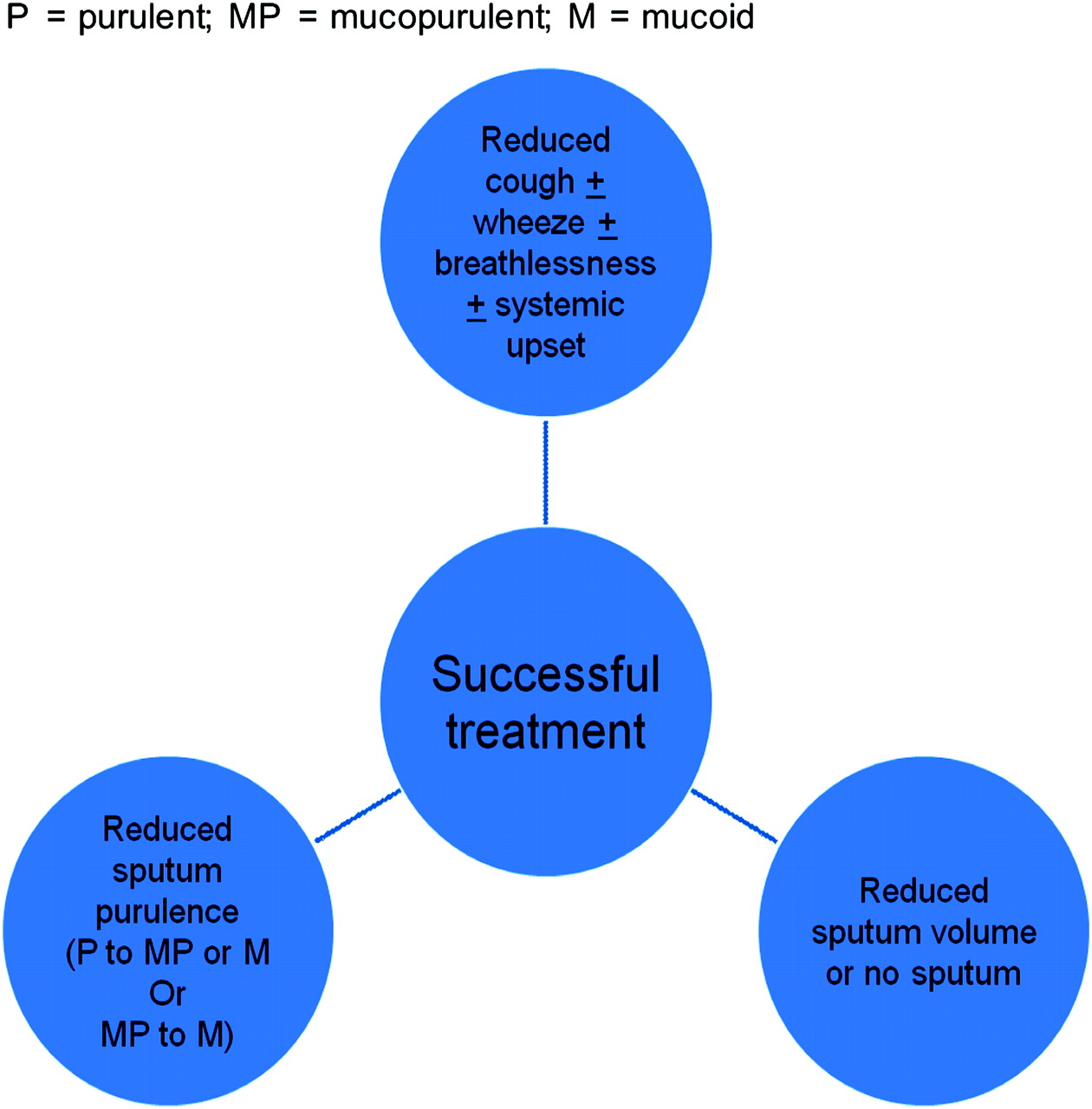
Definition of successful treatment of an exacerbation. P, purulent; MP, mucopurulent; M, mucoid.
Daily symptoms of cough and sputum production are frequent and patients with more severe bronchiectasis often expectorate mucopurulent or purulent sputum and culture respiratory pathogens when apparently clinically stable. This is more common in adults. The presence of mucopurulent or purulent sputum alone or the isolation of a pathogen alone is not necessarily an indication for antibiotic treatment.
Good practice points
The presence of mucopurulent or purulent sputum alone or the isolation of a pathogen alone is not necessarily indications for antibiotic treatment, particularly in adults.
Antibiotics should be given for exacerbations that present with an acute deterioration with worsening symptoms (cough, increased sputum volume or change of viscosity, increased sputum purulence with or without increasing wheeze, breathlessness, haemoptysis) and/or systemic upset.
Managing patients with exacerbations
Adults and children
Managing patients with exacerbations requires an assessment of severity of the exacerbation and decision about whether to treat the patient in the community or in hospital. This will depend on both patient factors and also on the experience and resources of the team managing the patient. For instance, support for domiciliary intravenous antibiotic therapy is not universal. Suggested criteria for inpatient treatment are shown in box 4. Suggested criteria for assessing patients treated in outpatient or inpatient settings are shown in box 5.
Box 4 Criteria for inpatient treatment of an exacerbation
Adults
Inpatient treatment recommendations if:
Unable to cope at home.
Development of cyanosis or confusion.
Breathlessness with respiratory rate ≥25/min.
Circulatory failure.
Respiratory failure.
Temperature ≥38°C.
Unable to take oral therapy.
Intravenous therapy required in patients with clinical failure after oral antibiotics (in hospitals with no facilities for domiciliary intravenous therapy).
Children
Inpatient treatment recommendations if:
Breathlessness with a raised respiratory rate and increased work of breathing.
Circulatory failure.
Respiratory failure.
Development of cyanosis.
Temperature ≥38°C.
Unable to take oral therapy.
Intravenous therapy required in patients with clinical failure after oral antibiotics (in hospitals with no facilities for domiciliary intravenous therapy).
Box 5 Assessment of patients with exacerbations of bronchiectasis
Adults
Outpatients
History.
Clinical examination.
Sputum for culture (preferably prior to commencement of antibiotics).
Review of previous sputum microbiology.
Inpatients
History.
Clinical examination.
Oxygen saturation on air.
Arterial blood gases if indicated.
ECG if indicated.
Chest x-ray.
Sputum for culture (preferably prior to commencement of antibiotics).
Blood culture if pyrexial ≥38°C.
Full blood count, urea and electrolytes and liver function tests.
Erythrocyte sedimentation rate or C-reactive protein (may be useful for diagnosing and monitoring exacerbations).
Review of previous sputum microbiology.
If feasible, 24 h sputum weight or volume.
Children
Outpatients
Sputum for culture if spontaneous sputum (preferably prior to commencement of antibiotics)
If spontaneous sputum is not available, throat/cough swabs, especially if regularly repeated, may provide inferred evidence of lower respiratory tract organisms.515 549 Caution should be taken in interpreting these cultures because many of the pathogens may also be normal upper airway commensals.
Induced sputum using hypertonic saline (3–5%) may be used to obtain lower respiratory tract samples for analysis.
Inpatients
In general history, clinical examination and oxygen saturation on air would be used to guide care at the onset of an inpatient stay.
Antibiotic choices would be made on the evidence of previous microbiology or to cover expected organisms in a child of that age.
Arterial blood gases are rarely justified, although capillary blood gas estimation of carbon dioxide may be used in a severe exacerbation.
Chest x-ray is warranted if children fail to respond to therapy or have unusual chest signs.
Children with systemic upset would warrant blood sampling (full blood count, urea and electrolytes, liver function tests, erythrocyte sedimentation rate or C-reactive protein).
In children failing to respond to initial therapy who are not sputum producers, induced sputum or bronchoalveolar lavage is warranted to identify infecting organisms.
Good practice points
Patients with an infective exacerbation of bronchiectasis should be assessed for the need for inpatient or outpatient treatment.
Patients treated for exacerbations should have appropriate assessments according to treatment setting.
Use of antibiotics
Which antibiotic regimen is recommended for exacerbations?
Adults
There are no randomised placebo-controlled trials in bronchiectasis. The studies of antibiotics in exacerbations of bronchiectasis are summarised in table AV in Appendix 2.236 237 246 368 369 424–431 The studies are heterogeneous and predominantly are in exacerbations treated in hospital. They indicate that a successful clinical outcome (figure 3) is achievable if there is high dosage targeted antibiotic therapy. High doses are often required to achieve the sputum becoming mucoid and bacterial clearance,368 369 424 426–428 430 although these goals are not always achievable. The duration of antibiotic therapy required needs further study. In one study the inflammatory response returned to normal within 7 days of antimicrobial therapy429 but symptomatic improvement has generally been seen in studies employing 10–14 days of treatment. Expert consensus is that 14 days should be recommended for all exacerbations. Further studies are needed to assess whether shorter regimens would suffice in exacerbations, particularly in patients with mild bronchiectasis.
The antibiotic choice is usually empirical and based on the likely microbial agent and perhaps informed by knowledge of previous sputum cultures in an individual. The antibiotic chosen should be based on local microbial patterns, sensitivities and cost. In those with mixed colonisation, an antibiotic should be chosen that will cover the organisms. Initial treatment will usually be with oral antibiotics with intravenous therapy reserved for those who fail to respond or who are particularly unwell. While exacerbations may be triggered by viral infections, there are no studies of the role of antiviral agents in exacerbations.
The common organisms associated with exacerbations of bronchiectasis and recommended antimicrobial agents are shown in table AI (Appendix 2). In the majority of patients the organism will be H influenzae and treatment with a β-lactam is appropriate (amoxicillin 500 mg three times daily for 14 days). Clinical improvement may be achieved with higher doses (eg, amoxicillin 1 g three times daily or 3 g twice daily) in patients who fail to respond to standard doses,236 368 and alternatives may need to be considered in the event of a β-lactamase-producing organism or penicillin sensitivity (table AI). Pseudomonas aeruginosa should be treated with an oral quinolone (ciprofloxacin 500–750 mg twice daily), although there is a significant chance of antibiotic resistance with poor clinical response after repeated courses. In addition, this class of antibiotic is associated with Clostridium difficile colitis, particularly in elderly patients.433 434 Patients with this organism often require intravenous therapy to achieve a clinical improvement.
The recommended route of antibiotic needs further study to address the optimal regimen. Previous studies showed that the combination of intravenous and inhaled antibiotics may have greater efficacy than intravenous therapy alone.369 427 430 In patients chronically colonised with P aeruginosa, however, the addition of nebulised tobramycin (300 mg twice daily) to high-dose oral ciprofloxacin (750 mg twice daily) for 14 days led to a greater reduction in microbial load at day 14 but there was no clinical benefit.432 Further studies are needed.
Recommendations (1−,236237246369424–427 3368428–434)
Before starting antibiotics, a sputum sample should be sent off for culture. [D]
Empirical antibiotics should be started whilst awaiting sputum microbiology. [D]
If there is no previous bacteriology, first-line treatment is amoxicillin 500 mg three times a day [B] or clarithromycin 500 mg twice daily (in patients that are penicillin-allergic) for 14 days. [C]
High-dose oral regimens (eg, amoxicillin 1 g three times a day or amoxicillin 3 g twice daily) may be needed in patients with severe bronchiectasis chronically colonised with H influenzae. [B]
Ciprofloxacin should be used in patients colonised with P aeruginosa with cautious use in elderly subjects. [B]
Previous sputum bacteriology results can be useful in deciding which antibiotic to use. Table AI highlights the recommended first-line and alternative treatments for the common bacterial pathogens implicated in exacerbations of bronchiectasis. [C]
Antibiotics can be modified subsequently once the pathogen is isolated only if there is no clinical improvement and the treatment should then be guided by antibiotic sensitivity results. [D]
Failure to respond to an antibiotic course should prompt a repeat sputum culture. [D]
Intravenous antibiotics should be considered when patients are particularly unwell, have resistant organisms or have failed to respond to oral therapy (this is most likely to apply to patients with P aeruginosa). [C]
There is no evidence to support the routine use of antiviral drugs in exacerbations. [D]
Children
There is no evidence available to help identify the most efficient antibiotic for use in paediatric bronchiectasis, nor evidence to help guide the optimal length of treatment required.
Good practice points
Previous sputum bacteriology results can be useful in deciding which antibiotic to use. Table AI highlights the first-line and alternative treatments for the common bacterial pathogens implicated in exacerbations of bronchiectasis (see BNF for Children for dosage; use doses for severe infection495).
Where possible, sputum (spontaneous or induced) or a cough swab should be obtained for culture prior to commencing antibiotics.
Empirical antibiotics can then be started while awaiting sputum microbiology.
In general, antibiotic courses for 14 days are standard. If there is no previous bacteriology, the first-line treatment is amoxicillin for 14 days or clarithromycin for 14 days in patients who are allergic to penicillin (see BNF for Children for dosage; use doses for severe infection495).
Children not responding to empirical antibiotic courses should have an organism identified by cough swab or later by induced sputum/bronchoalveolar lavage.
Intravenous antibiotics should be considered when patients are particularly unwell, have resistant organisms or have failed to respond to oral therapy (this is most likely to apply to patients with P aeruginosa).
When are combination (dual) antibiotic regimes required?
Adults and children
There is no evidence to recommend combination antibiotics in patients colonised with H influenzae, M catarrhalis, S aureus (methicillin-sensitive) and S pneumoniae. If there is more than one pathogen, an antibiotic should be selected that will cover both pathogens. If this is not feasible due to resistance patterns, combination antibiotics may be required.
In patients colonised with P aeruginosa there is controversy as to whether combination antibiotics are necessary. Studies in CF to date have shown similar efficacy with monotherapy as with combined antibiotics. Intravenous ceftazidime monotherapy was equally efficacious to intravenous ticarcillin/tobramycin in terms of clinical outcomes and reducing P aeruginosa colony counts.435 In another comparison, intravenous ceftazidime alone gave better lung function results than intravenous tobramycin and carbenicillin together.438 In another study intravenous azlocillin alone was compared with an intravenous tobramycin/azlocillin combination. Clinical outcomes were similar but the density of P aeruginosa fell more in the combination group.436 In patients with non-CF bronchiectasis chronically colonised with P aeruginosa the addition of nebulised tobramycin 300 mg twice daily to high-dose oral ciprofloxacin 750 mg twice daily for 14 days led to a greater reduction in microbial load at day 14, but there was no clinical benefit.432
A meta-analysis of single versus combination antibiotic therapy in CF did not show differences in response but was associated with an increase in the number of patients with resistant P aeruginosa at 2–8 weeks in patients on monotherapy.437 A consensus statement concluded that, with a susceptible strain, monotherapy may be as effective as combination treatment. Combination treatment was encouraged with a resistant strain and lowered the risk of developing further antibiotic resistance.439
Common practice for the intravenous antibiotic treatment of P aeruginosa is the combination of a third-generation cephalosporin such as ceftazidime with an aminoglycoside (usually gentamicin) for 14 days. Clinicians should be alert to the risk factors for aminoglycoside toxicity which are particularly relevant to patients with bronchiectasis (renal impairment, increasing age and long duration of treatment). Anecdotal reports of vestibular toxicity in older patients given gentamicin using a 7 mg/kg once daily regime suggest this is not appropriate for this group of patients (personal communication). Advice on the use of aminoglycosides is given in Appendix 1.
Recommendations (1+,432435436 1−,437438 4439)
Adults
Combination antibiotics are not required in patients colonised with H influenzae, M catarrhalis, S aureus (methicillin-sensitive) and S pneumoniae. [D]
If there is more than one pathogen, select an antibiotic that will cover both pathogens. If this is not feasible due to resistance patterns, combination antibiotics may be required. [D]
In patients who culture P aeruginosa that is sensitive to ciprofloxacin, monotherapy with oral ciprofloxacin can be used as first-line treatment (table AI). [B]
In patients who have not responded to oral ciprofloxacin, monotherapy with an antipseudomonal intravenous antibiotic should be considered (table AI). [D]
Combination antibiotics should be used for infections due to strains of P aeruginosa that are resistant to one or more antipseudomonal antibiotics (including ciprofloxacin) or if the clinician suspects the patient will require many subsequent antibiotic courses to reduce the development of drug resistance. [D]
Methicillin-resistant S aureus (MRSA) should be treated with two oral antibiotics or a single intravenous agent (see table AI). [D]
Intravenous aminoglycosides should only be used with appropriate and robust dosing and monitoring systems in place that have been agreed with local microbiologists and pharmacists (Appendix 1). [D]
Children
In children who culture P aeruginosa that is sensitive to ciprofloxacin, monotherapy with oral ciprofloxacin can be used (table AI). [B]
Those children whose sputum cultures yield pathogens with multiple resistant patterns should be considered for combination antibiotic therapy (in particular for P aeruginosa) (table AI). [D]
Identification of MRSA infection should prompt a dedicated eradication programme that in children may include a course of intravenous antibiotics, should oral antibiotics be unsuccessful (table AI). [D]
Do long-term oral antibiotics influence long-term outcome?
Adults
The aims with long-term antibiotic treatment are to improve symptoms, reduce the number of infective exacerbations and to improve health status. Patients chronically colonised with P aeruginosa have increased hospital admissions, worse quality of life and may have an accelerated decline in FEV1.233 483 There are sound theoretical reasons for trying to modulate the persisting airway inflammation that appears to perpetuate airway colonisation. By reducing microbial load and bacterial products in the airway, clearance of bacteria should be enhanced and allow the airway an opportunity to heal. Antibiotics, particularly those of the macrolide and quinolone groups, show potentially beneficial immunomodulatory effects on host inflammatory responses.496 497 These effects are evident at antibiotic concentrations below those required to kill infecting or colonising bacteria and some clinical experience has accumulated with low-dose long-term use of antibiotics, particularly macrolides, in the treatment of chronic airways diseases.498 499 Controlled clinical trials are lacking.
The studies using long-term oral antibiotics are summarised in table AVI in Appendix 2.244 247 353 367 374 440–446 From the MRC placebo-controlled trial in 1957, long-term twice weekly oxytetracycline over 1 year led to reduced sputum purulence, fewer days confined to bed and fewer days off work.440 Long-term tetracycline (≥3 months) compared with placebo led to less lower respiratory tract illness and of shorter duration.374 In 1988 an open labelled study assessed the effect of 4 months of amoxicillin (not placebo-controlled and with different regimens).367 This led to reduced airways inflammation with reduced elastase activity, less albumin protein leakage, improved patient well-being (from patient diary cards), reduced sputum volume and colour, improved breathlessness and improved PEF rates. In 1990 a 32-week study compared high-dose amoxicillin with placebo.443 This showed clinical improvement, reduction in 24 h sputum volume and fewer days confined to bed and away from work. There was no effect on exacerbation frequency but exacerbations were less severe.
Macrolides have been studied in bronchiectasis. A pilot study in 1999 compared 8 weeks of erythromycin with placebo.247 In this study 76% were chronically colonised with P aeruginosa. Erythromycin had no effect on proinflammatory cytokines, no impact on microbial load but an improvement in 24 h sputum volume and improved FEV1 and FVC. In 2004 an open labelled study (not placebo-controlled) evaluated the effect of long-term azithromycin (mean 20 months)446 and reported a reduction in chronic colonisation, improved symptoms and reduced exacerbation frequency. A further open labelled study in 2005 evaluated the effect of 6 months of treatment with azithromycin.445 The authors reported reduced 24 h sputum volume, improved well-being and reduced exacerbation frequency. There are potentially serious side effects with long-term macrolides500 and appropriate monitoring and follow-up are necessary.
A systematic review by Evans and colleagues in 2003 (carried out before the above studies) concluded that continuous antibiotics improved symptoms but had no effect on lung function or exacerbation frequency although there was a lack of randomised controlled studies with these endpoints.501 The effect of long-term continuous antibiotics on mortality is unknown. Further studies are needed.
As there is a high risk of resistance with continuous use of quinolone antibiotics in patients colonised with P aeruginosa, this class of drug should be avoided in this patient group.244 502 503
Recommendations (1+,374440–443 1−,247 3244353367444–446)
Patients having ≥3 exacerbations per year requiring antibiotic therapy or patients with fewer exacerbations that are causing significant morbidity should be considered for long-term antibiotics. [C]
In the first instance, high doses should not be used to minimise side effects. [C]
The antibiotic regimen should be determined by sputum microbiology when clinically stable (table AII). [D]
Long-term quinolones should not be used until further studies are available. [C]
Macrolides may have disease-modifying activity and preliminary data suggest the need for a large randomised controlled trial. [C]
Children
A long-term aim for some children is cure of the bronchiectasis. Up to 27% may have complete resolution of bronchiectasis within 24–48 months, with improvement in CT appearance in a further third of patients.504
Controlled clinical trials are lacking in bronchiectasis. Evidence is available for the long-term use of azithromycin in patients with CF-derived bronchiectasis505 506 and diffuse panbronchiolitis.507 It is not yet clear whether azithromycin may be expected to have a similar function in patients with non-CF bronchiectasis, with a different organism profile for lower respiratory tract infection. Roxithromycin assessed by randomised controlled trial in children with bronchiectasis improved airway responsiveness but not FEV1 (roxithromycin is, however, not currently available) (table AVI).347
Long-term antibiotics may aid healing and prevent exacerbations. Some centres provide long-term antibiotics while an assessment is made as to the rate of progression of bronchiectasis (repeating HRCT 24–48 months later). Co-trimoxazole, co-amoxiclav, clarithromycin or azithromycin are the most commonly used long-term prophylactic agents in children. There are no long-term studies to suggest preference of one over another antibiotic.
Good practice points
Consider long-term antibiotics in children with frequent symptoms or severe disease.
Assess rate of progression of bronchiectasis on long-term antibiotics (repeating HRCT 24–48 months later).
Where possible, long-term antibiotic regimens should be determined by respiratory microbiology.
Long-term use of oral quinolones should be avoided.
Do long-term nebulised antibiotics influence long-term outcome?
Adults
The aims with long-term treatment are to improve symptoms, reduce the number of infective exacerbations and to improve health status. Targeting antibiotics to the airways can deliver high doses of bactericidal drugs directly to the airway with little risk of systemic toxicity. The studies using long-term nebulised antibiotics are summarised in table AVII in Appendix 2.367 368 447–452
In 1985 an open labelled study (not placebo-controlled) investigated the effect of 4 months nebulised amoxicillin in patients who had relapsed despite treatment with high-dose oral amoxicillin.451 Patients treated with nebulised amoxicillin reported less sputum purulence, reduced sputum volume and increased PEF rates. In 1997 a 3-day randomised placebo-controlled study assessed the effect of inhaled gentamicin on neutrophil activity and mucus secretion.449 Inhaled gentamicin led to reduced sputum myeloperoxidase (a measure of neutrophil numbers), reduced microbial load, reduced sputum volume and improved Borg breathlessness score, improved PEF rates and 6 min walk test.
Subsequent studies assessed the efficacy of nebulised antibiotics in patients chronically colonised with P aeruginosa. In 1999 a study compared 1 year of treatment with nebulised ceftazidime and tobramycin versus symptomatic treatment in 15 patients.447 The actively treated group had fewer admissions and shorter stays in hospital but there was no difference in lung function, gas exchange or oral antibiotic usage at the end of 12 months. A larger randomised placebo-controlled study (74 patients) of shorter duration (6 weeks) was conducted in 2000.450 One month of treatment with nebulised tobramycin reduced sputum colonising load (a mean reduction of 4.5 log10 colony-forming units/ml), one-third of actively treated patients had P aeruginosa eradicated from their sputum and this was associated with a greater chance of improvement in their medical condition. There was no improvement in spirometry, no reduction in hospitalisation and some patients reported increased cough, wheeze, chest pain and breathlessness. There was only a minor change in the number of tobramycin-resistant strains of P aeruginosa. In 2005 a 6-month crossover randomised placebo-controlled trial (30 patients) was carried out. Nebulised tobramycin reduced the number of hospital admissions and hospital bed days (mean 0.15 and 2.05 days in the tobramycin group vs 0.75 and 12.65 days in the placebo group) and P aeruginosa density compared with placebo. There was no reduction in number of exacerbations, antibiotic use, improvement in pulmonary function or measures of health status.448 In 2005 an open labelled study was performed in 41 patients treated with three cycles of nebulised tobramycin (14 days treatment and 14 days off treatment). Treatment led to improved symptom scores, improved quality of life and eradication of P aeruginosa in 22%. About 5% developed resistance to tobramycin and 22% stopped treatment probably or possibly related to treatment due to cough, wheeze and breathlessness.452
Nebulised colistin is used in patients with bronchiectasis colonised with P aeruginosa. An uncontrolled study examining its efficacy in a mixed population of patients with COPD and bronchiectasis (the majority of whom had P aeruginosa) found an improvement in quality of life and slower decline in FEV1 with treatment.453
Recommendations (1+,447448 1−,449450 3367368451–453)
Patients having ≥3 exacerbations per year requiring antibiotic therapy or patients with fewer exacerbations that are causing significant morbidity should be considered for long-term nebulised antibiotics. [C]
In such patients, long-term nebulised antibiotics should be considered if chronically colonised with P aeruginosa (table AII). The choice of antibiotic should be guided by the antibiotic sensitivity results. Further studies are needed to address the optimal antibiotic choice and doses required. [C]
Research recommendation
Randomised controlled trials are needed to investigate the efficacy of nebulised antibiotics in patients with bronchiectasis chronically colonised with P aeruginosa and other organisms.
Children
Nebulised antibiotics may be considered in paediatric patients who experience frequent recurrent exacerbations (or deteriorating bronchiectasis) unaffected by long-term oral antibiotics or if colonised with P aeruginosa. Nebulised gentamicin (80 mg twice daily) in children with bronchiectasis is well tolerated and produces satisfactory drug levels in sputum (table AII).508 There is an extensive literature in CF using aerosolised antibiotics; for instance, 3-month periods of nebulised tobramycin (1 month on, 1 month off treatment repeated twice) improved FEV1 by 10% and decreased P aeruginosa density, use of intravenous antibiotics and hospital days in a large trial of 520 adult and paediatric patients with CF.509 Unlike the placebo group, there was an increase in the number of strains with minimum inhibitory concentration (MIC) for tobramycin >8 μg/ml, and it is for this reason that treatment is prescribed for 28 days followed by a drug holiday for the same period.
Good practice points
Long-term nebulised antibiotics are for children with frequent recurrent exacerbations (or deteriorating bronchiectasis) despite long-term oral antibiotics or if oral antibiotic therapy is not appropriate.
If a child with chronic P aeruginosa meets the criteria for long-term antibiotics, these should be considered; regimens are shown in table AII.
The choice of antibiotic should be guided by the antibiotic sensitivity results.
Research recommendations
Further studies are needed to address the optimal antibiotic choice and doses required.
Randomised controlled trials are needed to investigate the efficacy of nebulised antibiotics in children with bronchiectasis chronically colonised with organisms other than P aeruginosa.
Are rotational antibiotics recommended?
Adults and children
Policies for rotational use of antibiotics have been developed and implemented mainly in intensive care units and critical care facilities. The aim in these settings is to reduce the likelihood of the emergence and persistence of antibiotic-resistant strains within these hospital facilities, and the policies have met with some success.510–514 The extension of this concept to the long-term outpatient antibiotic treatment of individual patients with chronic lung diseases has some theoretical merit, but there are no suitable published studies to assess its value in this setting.
Good practice point
Rotating courses of antibiotics are not recommended routinely.
Should an attempt be made to eradicate organisms from the lower respiratory tract?
Adults and children
Some organisms, if able to colonise the lower respiratory tract in patients with bronchiectasis, can be difficult to treat. P aeruginosa often requires expensive and potentially toxic intravenous antibiotic therapy that is inconvenient and time-consuming for the patient; MRSA may be resistant to multiple antibiotics and has implications for infection control in the management of a patient with bronchiectasis in hospital. Colonisation with P aeruginosa is also associated with worse symptoms515 and quality of life scores233 and may lead to accelerated decline in FEV1.233 483 While there are no studies to guide practice following the first isolate of either organism, an attempt to eradicate seems pragmatic. Figures 4 and 5 outline strategies derived for the eradication of P aeruginosa from the sputum of patients with CF and individual clinicians will decide which and how aggressive a strategy to employ in the setting of non-CF bronchiectasis.

Eradication algorithm for Pseudomonas aeruginosa in adults. Attempt to eradicate with a 2-week course of oral ciprofloxacin (step 1). If step 1 fails, further regimens may be considered (step 2).

Eradication algorithm for Pseudomonas aeruginosa in children. Use doses according to the Children’s BNF. Use doses for severe infection.444 Attempt to eradicate with a 2-week course of oral ciprofloxacin (step 1). If step 1 fails, further regimens may be considered (step 2).
Good practice points
In patients who have P aeruginosa isolated for the first time, an attempt should be made to eradicate using 14 courses of oral ciprofloxacin (figures 4 and 5).
Failure to eradicate P aeruginosa with oral treatment may lead to consideration of intravenous and/or nebulised eradication therapy although there is currently insufficient evidence to recommend this (figures 4 and 5).
For patients in whom MRSA is isolated in the sputum, an attempt to eradicate the organism should be made with drug(s), dose and duration guided by local microbiological advice.
Antibiotic resistance
What is the impact of long-term antibiotics on antibiotic resistance?
Adults
The development of significant bacterial resistance in individual patients during prolonged treatment has occasionally been seen. During an 8-month trial of high-dose amoxicillin,443 this was not regarded as of any significance but was of more concern with long-term ciprofloxacin (≥90 days) where it was associated with clinical deterioration in 2 of 10 cases.244 There is a theoretical concern regarding the potential for prolonged antibiotic therapy to contribute to the emergence of antibiotic resistance in the community at large. However, when used for relatively small numbers of individual patients dispersed in the community, any impact on the overall resistance patterns of the common respiratory pathogens would be small and difficult to measure.
Recommendations (1+443, 3244)
Long-term antibiotics may result in antibiotic resistance in individual patients and alternative antibiotics should be chosen depending on sensitivity results. [D]
Long-term ciprofloxacin should not be used. [D]
Children
The development of significant bacterial resistance during prolonged treatment has occasionally been seen, but not to the same extent as with other chronic respiratory diseases such as CF. There is no large-scale published evidence on the emergence of antibiotic resistance patterns in children with bronchiectasis. There is a theoretical concern regarding the potential for prolonged antibiotic therapy to contribute to the emergence of antibiotic resistance. However, when used for relatively small numbers of individual patients dispersed in the community, any impact on the overall resistance patterns of the common respiratory pathogens would be small and difficult to measure.
Good practice point
Long-term antibiotics may result in antibiotic resistance in individual patients and alternative antibiotics should be chosen depending on sensitivity results.
Is there clinical relevance of in vitro antibiotic resistance patterns?
Adults and children
The prevalence of antimicrobial resistance in the common pathogens shows regional variation even within the UK, emphasising the significance of local antimicrobial susceptibility patterns. Overall, the rate of resistance to community-acquired respiratory pathogens was recently found to be low.454 The impact of resistance to penicillin in S pneumoniae is probably small in this setting. Satisfactory clinical outcomes have been reported following treatment of pneumonia caused by penicillin-resistant strains (MIC ≥2.0 μg/ml) with penicillins and cephalosporins.455 The prevalence of resistance in H influenzae in the UK is thought to be low and, as such, is not thought to have significant clinical impact at present.
By contrast, antimicrobial resistance among S aureus (eg, MRSA), P aeruginosa and Enterobacteriaceae (E coli, Klebsiella spp., Enterobacter spp.) presents significant potential problems, especially with the emergence of extended spectrum β-lactamases in the latter group. MRSA is resistant to all β-lactams and some common strains (eg, EMRSA-15, EMRSA-16) are frequently resistant to macrolides and ciprofloxacin. In children the range of antimicrobial agents available is further limited; for instance, tetracyclines which are useful alternatives in adults are contraindicated in children.
Recommendations (3454455)
Treatment should be guided on antibiotic sensitivity results but is often empirical based on previous sputum bacteriology. [D]
Some patients may respond to antibiotic treatment despite resistance to that drug in vitro. Antibiotics should only be changed if there is no clinical response. [D]
Section 6: Surgery, complications of bronchiectasis and management of advanced disease
Surgery for bronchiectasis
Is there a role for surgery in the management of patients with bronchiectasis?
Numerous studies have reported lung resection surgery for bronchiectasis in both children and adults with the principal aim of improving symptoms. Indications for surgery in the literature have included failure of medical/conservative therapy,460 464–467 518–521 recurrent respiratory infections and persistent sputum production,456 457 460 520–522 haemoptysis,460 464–467 518 523 524 chronic cough459 460 464 520 524 and persistent lung abscess.465–467 518 521
While lung resection surgery has not been subjected to randomised controlled trials, certain principles that seem to predict success emerge from the literature. First, the resection of bronchiectatic lung should be complete,458 461 464 465 518 520 525 526 so diseased lung must be localised463 465 518 and at least 10 segments should remain after surgery.527 There is no available information on contraindications, although it is unlikely that there are any absolute contraindications. Factors that appear to have an adverse outcome in some studies and so might be seen as relative contraindications include non-cylindrical disease,462 520 Pseudomonas grown on sputum culture,526 residual disease after resection and non-localised disease.458 461 463
Perioperative morbidity and mortality has been reported in many case series. The precise definition of morbidity varies, as does the reported incidence with complication rates ranging from 0%462 464 521 to >20%.518 520 523 The higher rates are in the older series and a morbidity rate of 10–19% is accepted by most authors.456 465 526 528 Perioperative mortality is low with all series reporting rates <5%460 465 518 519 521 and many with figures of 0%.458 466 467 520 524 526
With regard to long-term outcome, studies have reported many different variables with most concentrating on symptomatic improvement and subsequent admission rates. Improvement rates of 50–80%456 459 519–521 528 and >90%461 525 have been reported, although these studies do not formally control for a comparison group receiving best medical therapy.
Recommendations (3456–467)
Lung resection surgery may be considered in patients with localised disease in whom symptoms are not controlled by medical treatment. [D]
Patients undergoing surgery should have a review by a chest physician before referral. [D]
Massive haemoptysis
Haemoptysis is a rare but potentially life-threatening complication of bronchiectasis. There is no literature on the management or non-CF haemoptysis in children, with some studies involving adults with non-CF bronchiectasis. The principles of management follow those for CF; the first priority is to maintain the airway, optimise oxygenation and stabilise the haemodynamic status followed by bronchial artery embolisation or surgical intervention.472 Percutaneous bronchial artery embolisation has been shown to be a safe and effective method of controlling haemoptysis in both CF and non-CF populations.468–471 473
Recommendation (3468–473)
Bronchial artery embolisation and/or surgery is first-line therapy for the management of massive haemoptysis. [D]
Non-invasive ventilation (NIV)
There is little information on the short- or long-term role of NIV in the management of non-CF bronchiectasis with regard to physiological outcomes, survival and quality of life. A study of survival of 48 patients in the intensive care unit (ICU) where one of the interventions was NIV (n=13, 27%) found cumulative mortality of 19% (n=9) for a first admission to ICU for respiratory failure in patients with bilateral non-CF bronchiectasis and 40% (n=19) at 1 year.477 The actuarial survival rate at 1 year was 60%. Intubation requirement was associated with reduced survival in univariate analysis, suggesting that NIV may be advantageous. Multivariate analysis found that age >65 years and long-term oxygen therapy were independent factors predicting reduced survival. Survival was not reduced for those treated with long-term non-invasive positive pressure ventilation.
A study of the outcome of domiciliary NIV noted that the response to NIV in this group of patients (n=13) was disappointing.476 The patient group had more severe hypoxaemia and hypercapnoea at the start, suggesting that NIV was introduced later in the natural history of the disease than in previous studies. The authors concluded that the probability of continuing NIV after 2 years in the bronchiectasis group was <20% and most patients became increasingly more ventilator-dependent with time. However, all reported improvement in quality of sleep and in levels of daytime activity. Eight considered that the improvement in their respiratory status outweighed the discomfort due to NIV.476
A retrospective study of 16 patients with severe diffuse bronchiectasis investigating NIV as rescue therapy for nocturnal home use with daytime oxygen found a significant increase in FEV1 12 months after initiation of NIV. In patients alive after 24 months there was a significant decrease in the length of hospital admissions.475 This could be a reflection of the three deaths being the high intensity users. In a study which compared patients with bronchiectasis treated with NIV with those treated with long-term oxygen therapy only, differences were found between the two groups. In the year before NIV was commenced, mean±SD length of time in hospital for the NIV and long-term oxygen therapy groups was 48±55 and 5±8 days, respectively; the year following home NIV the length of time in hospital for the two groups was 10±31 and 9±16 days, respectively.474 In contrast, one study did not find a reduction in hospital days with the instigation of non-invasive positive pressure ventilation in patients with bronchiectasis.529
Recommendations (3474–477)
NIV can improve quality of life in some patients with chronic respiratory failure due to bronchiectasis. [D]
Evidence for survival benefit is lacking, although for some patients are successfully treated with NIV for significant lengths of time which may reduce hospitalisations. [D]
Lung transplantation
Is there a role for lung transplantation in advanced bronchiectasis?
Lung transplantation is available for end-stage cardiopulmonary disease in children and adults, although there is a paucity of literature in bronchiectasis specifically. A general guideline is to refer patients for an evaluation for lung transplantation if the FEV1 is <30% or if there is a rapid progressive respiratory deterioration despite optimal medical management.530 531 In patients with poor lung function, the following additional factors should lower the threshold for considering referral for transplantation assessment: massive haemoptysis, severe secondary pulmonary hypertension, ICU admissions or respiratory failure (particularly if requiring NIV). It should be noted that antibody deficiency is not an absolute contraindication to transplantation. The age of the patient predicts outcome following transplantation and discussion with a transplant centre is the best way to assess whether patients aged >60 years should be referred.
Good practice point
Appropriate patients should be referred for lung transplantation assessment.
Oxygen therapy
When should oxygen therapy be used?
Guidance on the appropriate use of oxygen in acute and chronic settings can be found in the BTS guideline for emergency oxygen use in adult patients.532
Appendix 1 Nebulised and intravenous antibiotics: a practical guide to administration
Nebulised antibiotics
How do you assess a patient for nebulised antibiotics?
Before a patient is commenced on regular nebulised antibiotics at home, a test dose assessment should be undertaken in hospital when the patient's condition is stable as bronchospasm can occur when nebulised antibiotics are administered.533 534 Any allergies should be noted before undertaking the test. Baseline spirometry should be performed at baseline (pre-dose) and then at 15 and 30 min after the test dose.535 536
Good practice points
A test dose should be given to identify problems with bronchospasm (box 6).
Alert the patient to the possibility of bronchospasm developing after commencement of regular dosing and advise cessation of the drug were this to occur.
Ensure patients have appropriate support once commenced on nebulised antibiotic.
Box 6 Protocol for test dose of a nebulised antibiotic
Explain the procedure to the patient, warning of possible side effects (cough, wheeze, chest tightness and breathlessness).
Ensure the dose of the antibiotic to be tested is prescribed and check for a history of sensitivity to the drug (which is a contraindication to administration).
Check the name, dose and expiry date of the test drug and all related diluents (where applicable).
Before starting the procedure, check availability of a spirometer and all necessary nebulisation equipment, together with a supply of salbutamol 2.5 mg nebules or metered dose inhaler with spacer.
Ensure all procedures for spirometry and nebulisation follow infection control recommendations; the nebuliser used for the test dose should be the one subsequently taken home by the patient.
Carry out spirometry at baseline, then at 15 and 30 min after the end of the test dose
If the FEV1 drops by <15% and <200 ml and the patient does not experience side effects, it is safe to give the nebulised antibiotic but, at follow-up visits, check there are no symptoms of bronchospasm related to the nebulised antibiotic.
If the FEV1 drops by >15% and >200 ml or if symptoms of bronchospasm occur, administer salbutamol by nebuliser or inhaler, repeating spirometry at 15 min intervals until it returns to baseline.
If bronchospasm has occurred, repeat the test on a separate day giving nebulised salbutamol (inhaled or nebulised) 10 min before the nebulised antibiotic. If the FEV1 drops by <15% and <200 ml, it is safe to continue the nebulised antibiotic but giving a β agonist prior to the nebulised antibiotic. If the FEV1 drops by >15% and >200 ml, consider an alternative formulation or drug.
If there are no side effects the patient may leave 30 min after the end of the test dose.
How do you ensure effective delivery of nebulised antibiotic therapy?
Effective therapy is determined by an efficient delivery system, the characteristics of the drug particle size, deposition pattern, dose and pharmacokinetics, the age of the patient, degree of airways narrowing, breathing patterns and adherence to treatment. All of these factors influence the efficacy of the therapy.535 537 A BTS guideline on current best practice for nebuliser treatment has been published.538
Good practice points
A chest physician should assess whether nebulised antibiotics are indicated.
A multidisciplinary team including a chest physician, physiotherapist and respiratory nurse should coordinate the care of the patient.
Reference should be made to BTS guidelines on nebuliser treatment.538
What nebuliser equipment should be used for nebulising antibiotics?
Compressors and nebuliser tested to British and European standards should be used. The British Standard BS7711 part 3: Respiratory therapy equipment specification for gas powered nebulisers for the delivery of drugs specifies that the minimum performance and safety requirements for gas powered nebulisers include:
Leakage not exceeding 5% of the maximum fill.
The nebuliser (or packaging) must be marked with the manufacturer's identity, lot number, recommended driving gas flow and the maximum filling level of the liquid container.
The respirable fraction of aerosol must be at least 50% at each of the recommended flows.
Suitability of the nebuliser for use with anaesthetic breathing systems and/or ventilators.
The nebuliser manufacturer must supply the following information:
A description of the intended use.
Suitability of the nebuliser for use with anaesthetic breathing systems and/or ventilators.
Minimum, maximum and recommended driving gas flows and the driving gas pressures corresponding to these.
The respirable output at minimum, maximum and recommended flows.
The distribution of aerosol particle size at the manufacturer's recommended driving gas flow.
The residual volume (by weight) left in the nebuliser at each driving gas flow.
Any contraindications to the use of the nebuliser.
Light, quiet, reliable and robust equipment that is easy to maintain is preferable. A breath-assisted venturi nebuliser (with a mouthpiece and a filter system) with a compressor producing a flow rate of 6 l/min is a suitable system.537
The compressor should be cleaned as appropriate and checked for safety and efficiency in accordance with the manufacturer's recommendations.539 This will usually entail washing the nebuliser chamber with warm soapy water. Nebulisation should take no longer than 10 min in order to ensure maximum compliance.539 Needles, syringes and sharps boxes should be provided.535
Good practice points
Equipment tested to British and European standards should be used.
A breath-assisted venturi nebuliser and filter system should be used with a compatible compressor producing a flow rate at 6 l/min and nebulisation should take no longer than 10 min in order to ensure maximum compliance.
Equipment should be cleaned and maintained according to manufacturers’ recommendations.
How do you reconstitute antibiotics for nebulisation?
Antibiotics are available as solutions or powder. How they are reconstituted will vary depending on the drug used. The manufacturer's instructions should be consulted. They are usually reconstituted as a solution using saline or water as a diluent to a volume of 3–4 ml.539 The commonly nebulised antibiotics and their preparation is shown in table AVIII (Appendix 2).
Good practice point
Antibiotics are usually reconstituted as a solution using saline or water as a diluent to a volume of 3–4 ml (more for higher doses).
How should the nebuliser equipment be cleaned and maintained?
Nebulisers may act as a source of bacterial contamination.539 The ideal standards and methods for cleaning nebulisers have not yet been well established. Patients should follow the manufacturer’s recommendations. In general, the nebuliser should be rinsed and thoroughly dried after every use.540
Good practice points
Hands should be washed prior to handling supplies and solution.
In general, the nebuliser should be rinsed and thoroughly dried after every use. Some nebulisers can be placed in a dishwasher. Most manufacturers recommend that the nebuliser is sterilised once a week either by using sterilising fluid or boiling the nebuliser (except tubing) in a clean pan for a full 10 min. All parts should be dry before re-assembly. Care should be taken to ensure the electrical compressor does not get wet.
Electrical compressors should be thoroughly cleaned in between patients and have the inlet filter changed according to the manufacturer's instructions (usually every 3 months).
Hospitals issuing nebuliser/compressor systems should arrange for their regular servicing in accordance with manufacturers’ recommendations.
Patients should be equipped with necessary spares and travel equipment where necessary.
A universal Code of Practice for the maintenance and re-use of equipment should be produced.
Do nebulised antibiotics pose a health risk to staff or relatives?
Concern has been expressed that medical personnel and/or relatives are at risk from exposure to nebulised antibiotics in the atmosphere. Occasionally, members of staff caring for patients using nebulised antibiotics have experienced cutaneous rashes and bronchoconstriction.539 It is generally recommended that the nebuliser be fitted with a filter on the expiratory port to prevent any environmental contamination536 and damage to patients’ property.
Good practice point
Filter systems (either filter or tubing) should be fitted on the expiratory port to prevent environmental contamination.
What advice should be given to patients about nebulised antibiotics?
Patients should be provided with advice to ensure that their equipment is used safely and efficiently. This will include details of assembling the equipment, preparation of the drug, use of the equipment, disposal of sharps and cleaning instructions.537 Contact numbers in case of breakdown are also helpful. Patients should be provided with training (including a practical demonstration) and clear written instructions in how to use and maintain the equipment.537
If drugs such as bronchodilators are also prescribed, the patient should be aware of which order to take the treatment to optimise the effect—that is, bronchodilators, physiotherapy followed by nebulised antibiotics.539
Potential side effects should be discussed including risk of bronchoconstriction at time of delivery, cutaneous rashes (although rare) and a sore mouth (due to Candida albicans infection, although the incidence is not known).539
Good practice points
Patients should be given instructions to ensure the equipment is used safely and efficiently.
Potential side effects should be discussed.
Intravenous antibiotics
When should home intravenous therapy be considered?
Evidence that intravenous antibiotics administered at home can be as effective as hospital treatment and cause minimum disruption to the patient's lifestyle comes from the literature on CF,541–543 although it seems reasonable to extrapolate this to patients with non-CF bronchiectasis. It is essential that patients be carefully selected. Prerequisites include: good visual acuity and manual dexterity to perform self-administration, adequate facilities in the home (clean environment, refrigeration and a telephone),541 reliable adherence to therapy, secure venous access, proper training and supervision.539 A variety of drugs may be prescribed (eg, ceftazidime, gentamicin, tobramycin and colistin). The dose will depend on the weight of the patient. However, large doses may be required similar to those given in CF (eg, ceftazidime 2 g three times daily for adult patients).
Good practice points
Home intravenous therapy can be given to suitable patients.
There must be a mechanism for checking antibiotic levels.
Where should the first dose of intravenous antibiotics be administered?
The first dose of intravenous antibiotics should be administered in hospital.539 Any drug allergy should be clearly documented and noted before any antibiotic is administered. In patients with CF the commonest reactions are the development of rashes.539
Good practice point
The first dose of intravenous antibiotics should be given in hospital.
How should drugs be administered?
As a number of drugs may be prescribed, the summary of product characteristics data sheet should be consulted to identify how drugs should be diluted for infusion/bolus administration.539 Drugs are generally diluted as much as possible; for example, aminoglycosides can be diluted with 15–20 ml sodium chloride and given slowly over 5 min or drugs may be given in an infusion over 30 min. Treatment should be made as patient-friendly as possible by using bolus doses and/or prepacked delivery devices. Where appropriate, once or twice daily doses should be prescribed.539
Good practice points
Drugs may be given via a slow bolus injection or infusion according to manufacturers’ instructions.
Treatment should be made as patient-friendly as possible by using bolus doses and/or prepacked delivery devices and, where appropriate, once or twice daily doses should be prescribed.
How should aminoglycosides be used in adults?
What advice should be given to the patient?
Advice should be given regarding how to prepare and administer their intravenous medication. This will include checking the medication labels for accuracy, learning aseptic technique (including good hand washing), recognising any side effects related to the medication and instructions in how to dispose of equipment such as sharps safely.541
Patients also have to learn about the particular type of intravenous catheter inserted for antibiotic therapy (eg, peripheral line). This includes potential complications that could occur, and appropriate flushing instructions. Written information including contact telephone numbers should be provided.
Good practice point
Written and verbal instructions should be given to the patient to ensure safe and efficient administration of their intravenous medication.
How should aminoglycosides be used?
Gentamicin can have substantial renal and ototoxicity. To avoid toxicity, therapeutic monitoring of serum levels is required. This document describes:
How to calculate a dose using ideal body weight (IBW).
How to monitor blood levels.
The following patients are at increased risk of toxicity and gentamicin should be used with caution: volume-depleted patients; patients with deteriorating renal function from any cause; patients on other nephrotoxic drugs such as non-steroidal anti-inflammatory agents, angiotensin-converting enzyme inhibitors or diuretics; and patients with actual weight substantially less than IBW.
To calculate gentamicin dose:
Before dosing gentamicin you will need a calculator and the following information:
Patient’s age
Patient’s height
Patient’s IBW (calculated from table below)
Determine creatinine clearance (CrCl) using the Cockcroft and Gault equation:
CrCl must be calculated using the above equation.
Estimated GFR [eGFR] from the patient’s biochemistry result is not appropriate.
If serum creatinine is <60 μmol/l, use 60 μmol/l.
| Female | |||||||||||||||||
| Height (ft) | 5′ | 5′1″ | 5′2″ | 5′3″ | 5′4″ | 5′5″ | 5′6″ | 5′7″ | 5′8″ | 5′9″ | 5′10″ | 5′11″ | 6′ | 6′1″ | 6′2″ | 6′3″ | 6′4″ |
| Height (cm) | 152 | 155 | 157 | 160 | 163 | 165 | 168 | 170 | 173 | 175 | 178 | 180 | 183 | 185 | 188 | 190 | 193 |
| IBW (kg) | 45.5 | 47.8 | 50.1 | 52.4 | 54.7 | 57 | 59.3 | 61.6 | 63.9 | 66.2 | 68.5 | 70.8 | 73.1 | 75.4 | 77.7 | 80 | 82.3 |
| Male | |||||||||||||||||
| Height (ft) | 5′ | 5′1″ | 5′2″ | 5′3″ | 5′4″ | 5′5″ | 5′6″ | 5′7″ | 5′8″ | 5′9″ | 5′10″ | 5′11″ | 6′ | 6′1″ | 6′2″ | 6′3″ | 6′4″ |
| Height (cm) | 152 | 155 | 157 | 160 | 163 | 165 | 168 | 170 | 173 | 175 | 178 | 180 | 183 | 185 | 188 | 190 | 193 |
| IBW (kg) | 50 | 52.3 | 54.6 | 56.9 | 59.2 | 61.5 | 63.8 | 66.1 | 68.4 | 70.7 | 73 | 75.3 | 77.6 | 79.9 | 82.2 | 84.5 | 86.8 |
Dose of gentamicin and frequency
Dose according to table above.
| Ideal body weight (kg) | |||||
| CrCl (ml/min) | 40–49 | 50–59 | 60–69 | 70–79 | >79 |
| 20–29 | 100 mg once daily | 100 mg once daily | 100 mg once daily | 160 mg every 48 h | 180 mg every 48 h |
| 30–39 | 120 mg once daily | 120 mg once daily | 140 mg once daily | 140 mg once daily | 160 mg once daily |
| 40–49 | 120 mg once daily | 140 mg once daily | 140 mg once daily | 160 mg once daily | 180 mg once daily |
| 50–59 | 100 mg twice daily | 140 mg once daily | 160 mg once daily | 180 mg once daily | 180 mg once daily |
| 60–69 | 120 mg twice daily | 140 mg twice daily | 140 mg twice daily | 180 mg once daily | 180 mg once daily |
| 70–79 | 140 mg twice daily | 140 mg twice daily | 160 mg twice daily | 180 mg once daily | 200 mg once daily |
| 80–89 | 140 mg twice daily | 160 mg twice daily | 160 mg twice daily | 160 mg twice daily | 180 mg twice daily |
| 90–99 | 160 mg twice daily | 160 mg twice daily | 180 mg twice daily | 180 mg twice daily | 180 mg twice daily |
| >99 | 160 mg twice daily | 180 mg twice daily | 200 mg twice daily | 200 mg twice daily | 200 mg twice daily |
This is designed to achieve peak concentrations of 7–10 mg/l and trough concentrations of <2 mg/l. The initial levels should be checked at the third dose. Check trough level immediately pre-dose and peak level 1 h post-dose. If the levels are within the therapeutic range, the gentamicin levels and urea and electrolytes should be checked every 3–4 days.
If the trough concentration is >2 mg/l, the drug should be withheld until the trough is <2 mg/l and the dosing interval should be increased.
If the peak level is >10 mg/l the dose should be reduced and if the peak level is <7 mg/l the dose should be increased. If the dose or dosing interval requires to be adjusted, levels should be checked at the third dose after the dose change.
In patients with renal impairment (CrCl <20 ml/min), gentamicin is best avoided.
What can be done if venous access is poor?
If poor venous access is a problem, this can be improved with the use of a long line or a totally implantable venous access device (TIVAD).539 Ports are made of titanium with a silicone septum. TIVADs allow the administration of antibiotic therapy for bronchiectasis (although other fluids can be given via this route). It is a closed system that is totally implanted subcutaneously. Insertion should be performed by a surgeon or radiologist experienced in placement of the device. Early complications can occur, such as bleeding, pneumothorax, nerve lesions or catheter misplacement. Wound and catheter infection (4–5%), thrombosis (3–3.5%), catheter fracture or disconnection (0.5%) and secondary dislocation (1.5–2%) of the catheter are the most important long-term complications.544 545
It is vital that the device is cared for by staff or patients who are fully trained and have expertise in the everyday care of such devices. When used properly complications are rare, although patients with diabetes mellitus need to pay particular attention to aseptic technique and blood sugar control.544 These devices need scrupulous care and regular (usually monthly) flushing with 10 ml of heparin (100 units/ml) using full aseptic technique. Infection or blockages are the most common complications.546 547 However, one small study suggested that routine flushing of Portacaths on a monthly basis is unnecessary and that flushing on alternate months only is sufficient; the possibility of extending the time interval between routine flushes even further is being investigated.548
The port should only be accessed with a special Huber needle. Ports can usually be used long term (up to 2000 punctures over ≥2 years) providing they are used correctly. A 10 ml or larger syringe should be used as smaller syringes exert high pressures and risk damaging the line. A pressure of 40 psi should not be exceeded.
Generally when antibiotics are being administered, the line should be flushed with saline, administer medication, saline, heparin. Always flush with saline in between antibiotics.
Good practice point
Only healthcare professionals trained in the use of long lines or totally implantable venous access systems should care for such devices.
Appendix
| (A) Adults | ||||
| Organism | Recommended first-line treatment | Length of treatment | Recommended second-line treatment | Length of treatment |
| Streptococcus pneumoniae | Amoxicillin 500 mg tds | 14 days | Clarithromycin 500 mg bd | 14 days |
| Haemophilus influenzae (β-lactamase negative) |
|
| Clarithromycin 500 mg bd or ciprofloxacin 500 mg bd or ceftriaxone 2 g od (IV) | 14 days |
| Haemophilus influenzae (β-lactamase positive) | Co-amoxiclav 625 mg tds | 14 days | Clarithromycin 500 mg bd or ciprofloxacin 500 mg bd or ceftriaxone 2 g od (IV) | 14 days |
| Moraxella catarrhalis | Co-amoxiclav 625 mg tds | 14 days | Ciprofloxacin 500 mg bd | 14 days |
| Staphylococcus aureus (MSSA) | Flucloxacillin 500 mg qds | 14 days | Clarithromycin 500 mg bd | 14 days |
| Staphylococcus aureus (MRSA): oral preparations | <50 kg: Rifampicin 450 mg od + trimethoprim 200 mg bd | 14 days | <50 kg: Rifampicin 450 mg od + doxycycline 200 mg od | 14 days |
| >50 kg:Rifampicin 600 mg + trimethoprim 200 mg bd | >50 kg: Rifampicin 600 mg + doxycycline 200 mg od | 14 days | ||
| Third-line: Linezolid 600 mg bd | 14 days | |||
| Staphylococcus aureus (MRSA): intravenous preparations | Vancomycin 1 g bd* (monitor serum levels and adjust dose accordingly) or teicoplanin 400 mg od | 14 days | Linezolid 600 mg bd | 14 days |
| Coliforms (eg, Klebsiella, enterobacter) | Oral ciprofloxacin 500 mg bd | 14 days | Intravenous ceftriaxone 2 g od | 14 days |
| Pseudomonas aeruginosa | Oral ciprofloxacin 500 mg bd (750 mg bd in more severe infections) | 14 days | Monotherapy: Intravenous ceftazidime 2 g tds or tazocin 4.5 g tds or aztreonam 2 g tds or meropenem 2 g tds | 14 days |
| Combination therapy: The above can be combined with gentamicin or tobramycin or colistin 2 MU tds (<60 kg, 50 000–75 000 units/kg daily in 3 divided doses) | 14 days | |||
| (B) Children (for doses consult BNF for Children and use doses for severe infection)495 | ||||
| Organism | Recommended first-line treatment | Length of treatment | Recommended second-line treatment | Length of treatment |
| Streptococcus pneumoniae | Amoxicillin | 14 days | Clarithromycin | 14 days |
| Haemophilus influenzae (β-lactamase negative) | Amoxicillin | 14 days | Clarithromycin or ceftriaxone (IV) | 14 days |
| Haemophilus influenzae (β-lactamase positive) | Co-amoxiclav | 14 days | Clarithromycin or ceftriaxone (IV) | 14 days |
| Moraxella catarrhalis | Co-amoxiclav | 14 days | Ciprofloxacin | 14 days |
| Staphylococcus aureus (MSSA) | Flucloxacillin | 14 days | Clarithromycin | 14 days |
| Staphylococcus aureus (MRSA): oral preparations | Rifampicin + trimethoprim | 14 days | Rifampicin + doxycycline | 14 days |
| Third-line: Linezolid | 14 days | |||
| Staphylococcus aureus (MRSA): intravenous preparations | Vancomycin or teicoplanin | 14 days | Linezolid | 14 days |
| Coliforms (eg, Klebsiella, enterobacter) | Oral ciprofloxacin | 14 days | Intravenous ceftriaxone | 14 days |
| Pseudomonas aeruginosa | Oral ciprofloxacin | 14 days | Monotherapy Intravenous ceftazidime or tazocin or aztreonam or meropenem | 14 days |
| Combination therapy: The above can be combined with gentamicin or tobramycin or colistin | 14 days | |||
Caution with aminoglycosides in pregnancy, renal failure, elderly patients or those on multiple other drugs.
↵* Elderly patients (>65 years): 500 mg vancomycin every 12 h or 1 g once daily (BNF 54, September 2007).500
bd, twice daily; IV, intravenous; od, once daily; qds, four times daily, tds, three times daily.
Common organisms associated with acute exacerbation of bronchiectasis and suggested antimicrobial agents
| (A) Adults | ||
| Organism | Recommended first-line treatment | Recommended second-line treatment |
| Streptococcus pneumoniae | Amoxicillin 500 mg bd | Clarithromycin 250 mg bd |
| Haemophilus influenzae (β-lactamase negative) | Amoxicillin 500 mg bd | Clarithromycin 250 mg bd |
| Haemophilus influenzae (β lactamase positive) | Co-amoxiclav 375 1 tablet tds | Clarithromycin 250 mg bd |
| Moraxella catarrhalis | Co-amoxiclav 375 1 tablet tds | Clarithromycin 250 mg bd |
| Staphylococcus aureus (MSSA) | Flucloxacillin 500 mg bd | Clarithromycin 250 mg bd |
| (B) Children and adults chronically colonised with Pseudomonas aeruginosa | |||
| Agent | Route | Adult dose | Frequency |
| Gentamicin | Nebulised | 80 mg | Twice daily |
| Tobramycin | Nebulised | 160 mg | Twice daily |
| Tobramycin (Tobi) | Nebulised | 300 mg | Twice daily |
| Colistin | Nebulised | 1–2 MU | Twice daily |
See BNF for Children (use doses for severe infection).495
Long-term oral antibiotic treatment
| Ref | Year | Country | N | Post-infection | Immune defect | CTD | ABPA | CF | Ciliary | IBD | Obstruction or foreign body | Aspiration or inhalation | Asthma | Congenital or airway abnormality | No cause identified |
| Children | |||||||||||||||
| 8 | 1994 | UK* | 41 | (32)† | 27 | NA‡ | 14 | 15% | 37% | ||||||
| 42 | 2001 | Turkey | 23 | 35 | 17 | 17 | 13 | 17 | 0% | ||||||
| 19 | 2003 | NZ | 66 | 25 | 12 | NA‡ | 10 | 1% | 50% | ||||||
| 23 | 2004 | UK§ | 93 | 30 | 26 | 0 | 1 | 3 | 1 | 8% | 18% | ||||
| 232 | 2005 | UK* | 136 | 4 | 34 | NA‡ | 15 | 18 | 4% | 25% | |||||
| All ages | |||||||||||||||
| 483 | 2000 | Italy | 49 | 6 | 4 | 2 | 0 | 4 | 2 | 10 | 2 | 4 | 6% | 55% | |
| Adults | |||||||||||||||
| 46 | 1995 | USA | 123 | 42 | 4 | 4 | 30% | ||||||||
| 22 | 2000 | UK* | 150 | 29 | 8 | 3 | 7 | 3 | 2 | 1 | 4 | 1% | 53% | ||
| 54 | 2003 | UK | 100 | 33 | 1 | 6 | 1 | 41% | |||||||
Data shown as percentages.
↵* Tertiary referral populations.
↵† Authors discounted prior pneumonia or pertussis in the absence of an immune defect as a possible cause of bronchiectasis.
↵‡ A diagnosis of cystic fibrosis was excluded prior to study entry.
↵§ Mixed secondary and tertiary referral population.
ABPA, allergic bronchopulmonary aspergillosis; CF, cystic fibrosis; CTD, connective tissue disease; IBD, inflammatory bowel disease.
Causes of bronchiectasis
| Year | Ref | Country | N | Age* | Method | Hi | Pa | Sa | Sp | Mc | Anaer | Spy | Asp | Myco | Non-pathogenic† | No growth |
| Children | ||||||||||||||||
| 1978 | 271 | USA | 38 | 0–16 | Sputum | 36‡ | 4 | 4 | ||||||||
| 2002 | 276 | Australia | 33 | 3.8 | BAL | 24 | 1 | <1 | <1 | <1 | 67 | |||||
| 2003 | 19 | New Zealand | 60 | 10 | Sputum | 68 | 2 | 12 | 6 | N/D | 8 | 4 | N/D | |||
| 2004 | 23 | UK | 93 | 0–16 | Sputum/BAL | 48 | 6 | 8 | 22 | 17 | N/D | 1 | 0 | N/D | ||
| 2005 | 232 | UK | 136 | 12.1 | Sputum/BAL | 39 | 11 | 4 | 22 | 2 | ? | <1 | ? | ? | ||
| Adults | ||||||||||||||||
| 1992 | 343 | Hong Kong | 91 | 50 | Sputum | N/D | N/D | N/D | N/D | N/D | N/D | N/D | N/D | 13§ | N/D | |
| 1995 | 46 | USA | 123 | 57 | Sputum | 30 | 31 | 7 | 11 | 2 | 2 | 5 | 23¶ | 23 | ||
| 1996 | 344 | UK | 135 | – | Sputum | N/D | 12 | N/D | N/D | N/D | N/D | N/D | N/D | N/D | N/D | |
| 1997 | 270 | Spain | 17 | 57 | BAL | 35 | 5 | N/D | N/D | 60 | 23 | |||||
| 2000 | 22 | UK | 150 | 53 | Sputum | 35 | 31 | 14 | 13 | 20 | N/D | 2 | N/D | 5 | ||
| 2001 | 268 | Spain | 49 | 57 | BAL | 26 | 20 | 2 | N/D | N/D | 28 | |||||
| 2002 | 145 | Thailand | 50 | 58 | Sputum | 14 | 20 | 6 | 4 | N/D | 6** | 36 | ||||
| 2002 | 342 | Spain | 42 | 58 | Sputum | 26 | 9 | 14 | 5 | N/D | 2 | 0 | 60 | |||
| 2002 | 342 | Spain | 59 | 58 | BAL | 32 | 10 | 3 | 7 | N/D | 0 | 32 | ||||
Data shown as percentages.
↵* Median age or range in children, mean in adult series.
↵† Non-pathogenic: non-pathogenic organisms such as Corynaebacterium, Neisseria spp, coagulase-negative Staphylococcus, β-haemolytic Streptococcus.
↵‡ Percentages may add up to more than 100% due to multiple isolates in one specimen/patient.
↵§ 11% Mycobacterium tuberculosis, 2% M avium, 1% M chelonae.
↵¶ 17% Mycobacterium avium intracellulare.
↵** 4% Mycobacterium kansasii, 2% M chelonae.
Anaer, anaerobe; Asp, Aspergillus; BAL, bronchoalveolar lavage; Hi, Haemophilus influenzae; Mc, Moraxella catarrhalis; Myco, mycobacteria; N/D, not done; Pa, Pseudomas aeruginosa; Sa, Staphylococcus aureus; Sp, Streptococcus pneumoniae; Spy, Streptococcus pyogenes.
Studies of lower respiratory tract microbiology in patients with bronchiectasis
| Author | Study | N | Study design | Duration | Baseline microbiology (main pathogens identified) | Principal outcome |
| Douglas, 1957424 | Randomised | 131 | Exacerbations of CB or BE that did not respond to 1 or 2 MU IM penicillin (5–14 days) (n=60) were randomised to oral chloramphenicol 2 g/day vs oral oxytetracycline 2 g/day | 5–7 days |
| Treatment with chloramphenicol was superior (78% led to mucoid sputum) compared with oxytetracycline (39% led to mucoid sputum) (p<0.01) |
| Pines, 1967426 | Randomised | 197 | Exacerbations of CB or BE that did not respond to recent antibiotic course were randomised to penicillin 2 MU IM (14 days) + streptomycin 0.5 g IM (7 days) vs IM cephaloridine 1 g bd vs IM cephaloridine 2 g bd vs IM cephaloridine 2 g tds | 14 days |
| Only 6 g cephaloridine was superior (56% led to mucoid sputum) to penicillin and streptomycin (34% led to mucoid sputum) (p=0.05) |
| Pines, 1970427 | Randomised | 81 |
| 7 days–2 months | All patients (purulent CB or BE) had chronic colonisation with P aeruginosa |
|
| Pines, 1974430 | Open label | 21 | Ticarcillin 1 g qds (if P aeruginosa 12–20 g ticarcillin/day + nebulised ticarcillin 0.5–1 g qds) | 7–10 days | 29% cultured P aeruginosa |
|
| Davies, 1983428 | Open label | 38 | IM ceftazidime 2 g bd 10 days vs IM ceftazidime 2 g tds 10 days vs IM ceftazidime 2 g bd 14 days | 10–14 days | 89% isolated P aeruginosa |
|
| Lam, 1986425 | Randomised | 32 | Oral ofloxacin 200 mg tds vs oral amoxicillin 1 g tds | 10 days | 56% isolated P aeruginosa or Klebsiella |
|
| Hill, 986368 | Open label* | 33 |
|
|
|
|
| Lam, 1989237 | Randomised | 41 | Oral ofloxacin 200 mg tds vs oral amoxicillin 1 g tds | 10 days | 32% isolated P aeruginosa or Klebsiella | Ofloxacin led to higher bacterial eradication (94% ofloxacin vs 45% amoxicillin) and a greater percentage had mucoid sputum (70% ofloxacin vs 38% amoxicillin, p<0.05) but had similar 3-month relapse rates |
| Ip, 1993429 | Open label | 12 | Oral ofloxacin 300 mg bd | 14 days | 75% had pathogenic organisms, of which 67% had P aeruginosa |
|
| Tag El-Din, 1994369 | Randomised | 27 | Depending on sensitivities IV treatment 7 days vs IV treatment 7 days + inhaled amikacin or ceftazidime or gentamicin or tobramycin 7 days | 7 days | ∼62% cultured P aeruginosa, Klebsiella or E coli | Combination of inhaled + IV antibiotics led to improved FEV1, FVC and PEFR and >6-fold reduction in sputum volume compared with IV therapy alone (p<0.001) |
| Chan, 1996236 | Randomised | 42 | Oral ciprofloxacin 500 mg bd vs oral amoxicillin 1 g tds | 7 days | 41% isolated Pseudomonas spp. | Ciprofloxacin led to higher bacterial clearance (100% ciprofloxacin vs 25% amoxicillin) and improved sputum purulence (95% ciprofloxacin vs 55% amoxicillin) and lower sputum volume (p<0.05) |
| Tsang, 1999246 | Randomised | 35 | Oral levofloxacin 300 mg bd vs ceftazidime 1 g tds IV | 10 days | 29% isolated P aeruginosa, 41% no pathogens | Similar bacterial eradication rates (∼70%) and clinical outcomes (cough, sputum purulence, breathlessness) |
| Darley, 2000431 | Open label* | 9 | IV meropenem 3 g/day | 7–10 days | 67% had pathogenic organisms, of which 67% were P aeruginosa | 83% no pathogenic organisms at end of treatment |
↵* If treated as outpatients.
CB, chronic bronchitis; bd, twice daily; BE, bronchiectasis; FEV1, forced expiratory volume in 1 s; FVC, forced vital capacity; IM, intramuscular; IV, intravenous; PEFR, peak exploratory flow rate; tds; three times daily.
Antibiotic studies for exacerbations treated in hospital: adults
| (A) Adults | ||||||
| Author | Study | N | Study design | Duration | Baseline microbiology | Principal outcome |
| MRC, 1957440 | Randomised | 122 |
| 1 year, 2 days per week | None recorded |
|
| Cherniack, 1959374 | Randomised | 67 |
| ≥3 months (3–22) |
|
|
| Dowling, 1960441 | Randomised | 89 |
| ≥3 months (3–31) |
|
|
| Sobel, 1962442 | Randomised | 90 |
| Data up to 5 years but data up to 36 months in all groups | None available |
|
| Stockley, 1984444 | Open label | 15 | Study to assess effect of antibiotic treatment (amoxicillin 250 mg tds) on sputum elastase in stable bronchiectasis patients who normally produce purulent phlegm | 14 days |
|
|
| In non responders a further 2-week antibiotic course was given | 14 days | |||||
| Hill, 1986353 | Open label | 18 |
| 14 days | None recorded | ∼5% increase in FEV1, FVC, VC and TLC (p<0.05) |
| 10 patients received 4 months of amoxicillin and all had mucoid phlegm at the end of treatment | 4 months | Only increase in FVC (p<0.05) | ||||
| Hill, 1988367 | Open label | 10 |
| 16 weeks |
|
|
| Currie, 1990443 | Randomised | 38 | 3 g twice daily oral amoxicillin vs placebo | 32 weeks | 24% had P aeruginosa |
|
| Rayner, 1994244 | Retrospective | 10 | Study to assess efficacy and safety of long-term ciprofloxacin (500–1500 mg/day) | ≥90 days |
|
|
| Tsang, 1999247 | Randomised | 21 |
| 8 weeks |
|
|
| Davies, 2004446 | Open label | 39 | Study to assess the impact of long-term azithromycin | 20±10 months |
|
|
| Cymbala, 2005445 | Open label | 12 | Study to assess long-term impact of long-term azithromycin 500 mg twice weekly | 6 months | None recorded |
|
| (B) Children | ||||||
| Koh, 1997347 | Randomised | 25 | Effect of roxithromycin for 12 weeks on airway responsiveness in children (<16 years) with bronchiectasis: a double blind, placebo-controlled study | 12 weeks | Not stated |
|
bd, twice daily; FEV1, forced expiratory volume in 1 s; FVC, forced vital capacity; IL, interleukin; IM, intramuscular; IV, intravenous; LTB4, leukotriene B4; PD20, dose required to provoke a fall in FEV1 of 20%; PEF, peak expiratory flow; tds, three times daily; TLC, total lung capacity; TNFα, tumour necrosis factor α; VC, vital capacity.
Oral antibiotic studies used in stable bronchiectasis
| (A) Adults | ||||||
| Author | Study | N | Study design | Duration | Baseline microbiology | Principal outcome |
| Stockley, 1985451 | Open label | 6 | Study to assess the impact of 4 months nebulised amoxicillin in those who relapsed following amoxicillin 3 g bd | 4 months |
|
|
| Hill et al, 1986368 | Open label | 33 | Antibiotics given for exacerbations or in stable patients with intermittent mucopurulent/purulent phlegm or in patients with persistent purulent phlegm |
|
| |
| 1. Amoxicillin 250 mg tds and if failed to respond | 14 days | |||||
| 2. Amoxicillin 3 g bd and if failed to respond | 14 days | |||||
| 3. Nebulised amoxicillin 500 mg bd | 4 months | |||||
| Hill, 1988367 | Open label | 10 |
| 16 weeks |
|
|
| Lin, 1997449 | Randomised | 16 |
| 3 days | Not recorded |
|
| Orriols, 1999447 | Randomised | 15 |
| 1 year | 100% P aeruginosa |
|
| Barker, 2000450 | Randomised | 74 |
| 4 weeks on treatment, then 2 weeks off treatment | 100% P aeruginosa |
|
| Drobnic, 2005448 | Randomised | 30 |
| 6 months | 100% P aeruginosa |
|
| Scheinberg, 2005452 | Open label | 41 |
| 3 cycles of14 days on treatment and 14 days off treatment | 100% P aeruginosa |
|
| Steinfort, 2007453 | Open label | 18 | Nebulised colistin 30 mg in 18 patients (14 bronchiectasis, 4 COPD); majority with P aeruginosa | Mean 41 months | 78% P aeruginosa |
|
| (B) Children | ||||||
| Twiss, 2005508 | Open label safety study | 10 |
| Single dosing study |
|
|
bd, twice daily; COPF, chronic obstructive pulmonary disease; FEV1, forced expiratory volume in 1 s; FVC, forced vital capacity; PEF, peak expiratory flow; tds, three times daily.
Studies of nebulised antibiotics in patients with stable bronchiectasis
| Drug and formulation | Dose | Sodium chloride 0.9% (ml) | Sterile water (ml) | Total volume (ml) | Daily dose |
| Colistin539 (Colomycin)† | 1–2 MU |
|
|
|
|
| Colistin (Promixin)‡ | 1 MU |
|
|
|
|
| Gentamicin 40 mg/ml sol508 | 80 mg | 2 | – | 4 | Adults 160 mg bd |
| Tobramycin 40 mg/ml sol447 539 | 80 mg160 mg |
|
|
| Adults 80–160 mg bd |
| Tobramycin (Tobi) solution | 300 mg | – | – | 5 | Adults and children** 300 mg bd |
| Amoxicillin 500 mg dry powder451 | 500 mg | – | 5 | 5 | Adults 500 mg bd |
↵* Note the nebulised drugs in this table are not licensed by any manufacturer for use in non-cystic fibrosis bronchiectasis.
↵† Manufacturer states mixing with 0.9% saline is preferred with sterile water or 50% water/saline being alternatives.
↵‡ Mix with either sterile water or 50% sterile water/50% 0.9% saline; for use only in iNEB AAD nebuliser.
↵§ If age <2 years 500 000 units bd.
↵¶ Age ≥2 years.
↵** Age ≥6 years.
bd, twice daily.
Nebulised antibiotics*
Appendix 3 Audit criteria and research questions
Audit criteria
All patients should be assessed for underlying cause(s).
Sputum microbiology should be checked prior to antibiotics being given for exacerbations.
Summary of research recommendations
Further studies are required to establish the link between COPD and bronchiectasis.
Further studies in other connective tissue diseases are indicated.
Use of carbocysteine in bronchiectasis should be the subject of a randomised control trial to establish its clinical efficacy.
Mannitol should be investigated further in a randomised controlled trial.
A large randomised controlled trial is required to assess the role of inhaled corticosteroids in bronchiectasis.
Randomised controlled trials are needed to investigate the efficacy of nebulised antibiotics in patients with bronchiectasis chronically colonised with P aeruginosa and other organisms.
Further studies are needed to address the optimal antibiotic choice and doses required.
Randomised controlled trials are needed to investigate the efficacy of nebulised antibiotics in children with bronchiectasis chronically colonised with organisms other than P aeruginosa.
Further research is needed to investigate the efficacy of all the airway clearance techniques in non-CF bronchiectasis, particularly positive expiratory pressure, RC-Cornet, autogenic drainage and high-frequency chest wall oscillation.
Research questions
Are mucolytics (carbocysteine, mecysteine) effective in improving symptoms or exacerbation frequency?
Do strategies to eradicate P aeruginosa affect clinical outcome?
Do macrolides affect long-term outcome (symptoms, lung function, exacerbations, treatment requirements, quality of life)?—need for randomised controlled trial.
Do long-term oral or nebulised antibiotics improve outcome in children (symptoms, exacerbations, quality of life)?
Do long-term nebulised antipseudomonal antibiotics affect outcome in adults or children?
Does rotating different long-term oral antibiotics have any benefit (outcomes and antibiotic resistance)?
What is the outcome after lung transplantation in patients with bronchiectasis?
Appendix 4 Contributors
Steering Group
Dr Mark C Pasteur (Chairman), Dr Diana Bilton, Dr Adam T Hill
Working Group 1 (Introduction, Clinical Assessment, Investigations)
Introduction: Professor Robert A Stockley; Adult physicians: Drs Robert Wilson and Mark C Pasteur; Immunologist: Dr Richard Herriot; Radiologist: Professor David M Hansell; Paediatrician: Professor Andrew Bush; General practitioner: Dr Charles Cornford; Patient representative: Lorna Willcox.
Working Group 2 (Management, Airways clearance, Adjunctive treatments, Surgery)
Adult physician: Dr Diana Bilton; Surgeon: Mr G Wyn Parry; Paediatrician: Dr Samantha Sonnappa (assisted by Dr Colin Wallis); Specialist nurse: Jane French; Physiotherapists: Frances Sinfield, Alex Harvey (assisted by Julia Bott and Jennifer Pryor); General practitioner: Dr Charles Cornford; Patient representative: Lorna Willcox.
Working Group 3 (Antibiotics)
Adult physicians: Drs Adam T Hill and Mike Greenstone; Paediatricians: Drs Steven Cunningham and David A Spencer; Microbiologists: Drs Xavier Emmanuel and Pota Kalima; Specialist nurse: Karen Heslop; General practitioner: Dr Charles Cornford; Patient representative: Lorna Willcox.
External peer reviewers
Professor Stuart Elborn (Royal Belfast Hospital, Belfast); Dr Siobhán Carr (Barts and The London Children's Hospital, London).
Library services and literature searches
Bridget Cole (Head Librarian, Norfolk and Norwich University Hospital NHS Foundation Trust).
References
- ↵
- ↵
- ↵
- ↵
- ↵
- ↵
- ↵
- ↵
- ↵
- ↵
- ↵
- ↵
- ↵
- ↵
- ↵
- ↵
- ↵
- ↵
- ↵
- ↵
- ↵
- ↵
- ↵
- ↵
- ↵
- ↵
- ↵
- ↵
- ↵
- ↵
- ↵
- ↵
- ↵
- ↵
- ↵
- ↵
- ↵
- ↵
- ↵
- ↵
- ↵
- ↵
- ↵
- ↵
- ↵
- ↵
- ↵
- ↵
- ↵
- ↵
- ↵
- ↵
- ↵
- ↵
- ↵
- ↵
- ↵
- ↵
- ↵
- ↵
- ↵
- ↵
- ↵
- ↵
- ↵
- ↵
- ↵
- ↵
- ↵
- ↵
- ↵
- ↵
- ↵
- ↵
- ↵
- ↵
- ↵
- ↵
- ↵
- ↵
- ↵
- ↵
- ↵
- ↵
- ↵
- ↵
- ↵
- ↵
- ↵
- ↵
- ↵
- ↵
- ↵
- ↵
- ↵
- ↵
- ↵
- ↵
- ↵
- ↵
- ↵
- ↵
- ↵
- ↵
- ↵
- ↵
- ↵
- ↵
- ↵
- ↵
- ↵
- ↵
- ↵
- ↵
- ↵
- ↵
- ↵
- ↵
- ↵
- ↵
- ↵
- ↵
- ↵
- ↵
- ↵
- ↵
- ↵
- ↵
- ↵
- ↵
- ↵
- ↵
- ↵
- ↵
- ↵
- ↵
- ↵
- ↵
- ↵
- ↵
- ↵
- ↵
- ↵
- ↵
- ↵
- ↵
- ↵
- ↵
- ↵
- ↵
- ↵
- ↵
- ↵
- ↵
- ↵
- ↵
- ↵
- ↵
- ↵
- ↵
- ↵
- ↵
- ↵
- ↵
- ↵
- ↵
- ↵
- ↵
- ↵
- ↵
- ↵
- ↵
- ↵
- ↵
- ↵
- ↵
- ↵
- ↵
- ↵
- ↵
- ↵
- ↵
- ↵
- ↵
- ↵
- ↵
- ↵
- ↵
- ↵
- ↵
- ↵
- ↵
- ↵
- ↵
- ↵
- ↵
- ↵
- ↵
- ↵
- ↵
- ↵
- ↵
- ↵
- ↵
- ↵
- ↵
- ↵
- ↵
- ↵
- ↵
- ↵
- ↵
- ↵
- ↵
- ↵
- ↵
- ↵
- ↵
- ↵
- ↵
- ↵
- ↵
- ↵
- ↵
- ↵
- ↵
- ↵
- ↵
- ↵
- ↵
- ↵
- ↵
- ↵
- ↵
- ↵
- ↵
- ↵
- ↵
- ↵
- ↵
- ↵
- ↵
- ↵
- ↵
- ↵
- ↵
- ↵
- ↵
- ↵
- ↵
- ↵
- ↵
- ↵
- ↵
- ↵
- ↵
- ↵
- ↵
- ↵
- ↵
- ↵
- ↵
- ↵
- ↵
- ↵
- ↵
- ↵
- ↵
- ↵
- ↵
- ↵
- ↵
- ↵
- ↵
- ↵
- ↵
- ↵
- ↵
- ↵
- ↵
- ↵
- ↵
- ↵
- ↵
- ↵
- ↵
- ↵
- ↵
- ↵
- ↵
- ↵
- ↵
- ↵
- ↵
- ↵
- ↵
- ↵
- ↵
- ↵
- ↵
- ↵
- ↵
- ↵
- ↵
- ↵
- ↵
- ↵
- ↵
- ↵
- ↵
- ↵
- ↵
- ↵
- ↵
- ↵
- ↵
- ↵
- ↵
- ↵
- ↵
- ↵
- ↵
- ↵
- ↵
- ↵
- ↵
- ↵
- ↵
- ↵
- ↵
- ↵
- ↵
- ↵
- ↵
- ↵
- ↵
- ↵
- ↵
- ↵
- ↵
- ↵
- ↵
- ↵
- ↵
- ↵
- ↵
- ↵
- ↵
- ↵
- ↵
- ↵
- ↵
- ↵
- ↵
- ↵
- ↵
- ↵
- ↵
- ↵
- ↵
- ↵
- ↵
- ↵
- ↵
- ↵
- ↵
- ↵
- ↵
- ↵
- ↵
- ↵
- ↵
- ↵
- ↵
- ↵
- ↵
- ↵
- ↵
- ↵
- ↵
- ↵
- ↵
- ↵
- ↵
- ↵
- ↵
- ↵
- ↵
- ↵
- ↵
- ↵
- ↵
- ↵
- ↵
- ↵
- ↵
- ↵
- ↵
- ↵
- ↵
- ↵
- ↵
- ↵
- ↵
- ↵
- ↵
- ↵
- ↵
- ↵
- ↵
- ↵
- ↵
- ↵
- ↵
- ↵
- ↵
- ↵
- ↵
- ↵
- ↵
- ↵
- ↵
- ↵
- ↵
- ↵
- ↵
- ↵
- ↵
- ↵
- ↵
- ↵
- ↵
- ↵
- ↵
- ↵
- ↵
- ↵
- ↵
- ↵
- ↵
- ↵
- ↵
- ↵
- ↵
- ↵
- ↵
- ↵
- ↵
- ↵
- ↵
- ↵
- ↵
- ↵
- ↵
- ↵
- ↵
- ↵
- ↵
- ↵
- ↵
- ↵
- ↵
- ↵
- ↵
- ↵
- ↵
- ↵
- ↵
- ↵
- ↵
- ↵
- ↵
- ↵
- ↵
- ↵
- ↵
- ↵
- ↵
- ↵
- ↵
- ↵
- ↵
- ↵
- ↵
- ↵
- ↵
- ↵
- ↵
- ↵
- ↵
- ↵
- ↵
- ↵
- ↵
- ↵
- ↵
- ↵
- ↵
- ↵
- ↵
- ↵
- ↵
- ↵
- ↵
- ↵
- ↵
- ↵
- ↵
- ↵
- ↵
- ↵
- ↵
- ↵
- ↵
- ↵
- ↵
- ↵
- ↵
- ↵
- ↵
- ↵
- ↵
- ↵
- ↵
- ↵
- ↵
- ↵
- ↵
- ↵
- ↵
- ↵
- ↵
- ↵
- ↵
- ↵
- ↵
- ↵
- ↵
- ↵
- ↵
- ↵
- ↵
- ↵
- ↵
- ↵
- ↵
- ↵
- ↵
- ↵
Footnotes
Competing interests DB has undertaken consultancy for Transave and Aradigm. MCP and ATH have no competing interests.
Provenance and peer review Not commissioned; not externally peer reviewed.