 

Main menu

  • Home
  • Content
    • Current Issue
    • Editor's Commentary
    • Archives
    • Most-Read Papers of 2022
  • Authors
    • Author Guidelines
    • Submit a Manuscript
  • Reviewers
    • Reviewer Information
    • Create Reviewer Account
    • Reviewer Guidelines: Original Research
    • Reviewer Guidelines: Reviews
    • Appreciation of Reviewers
  • CRCE
    • Through the Journal
    • JournalCasts
    • AARC University
    • PowerPoint Template
  • Open Forum
    • 2023 Call for Abstracts
    • 2022 Abstracts
    • Previous Open Forums
  • Podcast
    • English
    • Español
    • Portugûes
    • 国语
  • Videos
    • Video Abstracts
    • Author Interviews
    • Highlighted Articles
    • The Journal

User menu

  • Subscribe
  • My alerts
  • Log in

Search

  • Advanced search
American Association for Respiratory Care
  • Subscribe
  • My alerts
  • Log in
American Association for Respiratory Care

Advanced Search

  • Home
  • Content
    • Current Issue
    • Editor's Commentary
    • Archives
    • Most-Read Papers of 2022
  • Authors
    • Author Guidelines
    • Submit a Manuscript
  • Reviewers
    • Reviewer Information
    • Create Reviewer Account
    • Reviewer Guidelines: Original Research
    • Reviewer Guidelines: Reviews
    • Appreciation of Reviewers
  • CRCE
    • Through the Journal
    • JournalCasts
    • AARC University
    • PowerPoint Template
  • Open Forum
    • 2023 Call for Abstracts
    • 2022 Abstracts
    • Previous Open Forums
  • Podcast
    • English
    • Español
    • Portugûes
    • 国语
  • Videos
    • Video Abstracts
    • Author Interviews
    • Highlighted Articles
    • The Journal
  • Twitter
  • Facebook
  • YouTube
Case ReportCase Reports

Congenital Pulmonary Airway Malformation: A Case Report and Review of the Literature

Ryan J McDonough, Alexander S Niven and Keith A Havenstrite
Respiratory Care February 2012, 57 (2) 302-306; DOI: https://doi.org/10.4187/respcare.00727
Ryan J McDonough
Department of Medicine, Madigan Army Medical Center, Tacoma, Washington.
  • Find this author on Google Scholar
  • Find this author on PubMed
  • Search for this author on this site
  • For correspondence: [email protected]
Alexander S Niven
Department of Medicine, Madigan Army Medical Center, Tacoma, Washington.
  • Find this author on Google Scholar
  • Find this author on PubMed
  • Search for this author on this site
Keith A Havenstrite
Department of Medicine, Madigan Army Medical Center, Tacoma, Washington.
  • Find this author on Google Scholar
  • Find this author on PubMed
  • Search for this author on this site
  • Article
  • Figures & Data
  • References
  • Info & Metrics
  • PDF
Loading

Abstract

Congenital parenchymal lung malformations have an estimated incidence at 1:25,000–1:35,000 births. We present a case of this rare congenital abnormality in a 38 year-old male, review the current literature with discussion of proposed causes, malignant potential, and management strategies. A 38-year-old white male presented with a 4-day history of chronic stable hemoptysis. Social history was notable for a 50-pack-year active smoking history and remote heavy alcohol consumption. Physical examination was normal. Chest radiograph revealed an ill defined right lower lobe infiltrate. Chest computed tomography demonstrated an irregular, thin-walled, cystic lesion with adjacent nodularity and calcifications. The patient received a right lower lobectomy. Pathologic specimen demonstrated a 10-cm, mostly thin-walled cyst with features suggestive of a congenital cyst adenomatoid malformation and areas of adenocarcinoma (mixed subtype with acinar and bronchioloalveolar patterns). Congenital cyst adenomatoid malformations have recently been renamed as congenital pulmonary airway malformations and are the most common type of congenital parenchymal lung malformations. Individuals typically present with recurrent pulmonary infections, pneumothorax, or hemoptysis. The development is controversial but believed to be a result of arrested development of the fetal bronchial tree during the sixth and seventh week of fetal development. Defects in thyroid transcription factor 1 have also been proposed. With the increasing use and image resolution of ultrasound in modern obstetric practice, congenital pulmonary airway malformations rarely go undetected into adulthood. Management remains controversial; however, most authors agree with early surgical excision.

  • congenital cystic adenomatoid malformation
  • CCAM
  • congenital lobar hyperinflation
  • bronchogenic cyst
  • lobar sequestration
  • hemoptysis

Introduction

Congenital parenchymal lung malformations are uncommon yet well described fetal anomalies, primarily identified in the pediatric population. They incorporate a spectrum of interrelated abnormalities that include congenital lobar hyperinflation, bronchogenic cyst, congenital cystic adenomatoid malformation (CCAM), and lobar sequestration.1 We present a case of hemoptysis in a 38-year-old male with CCAM and review the literature relating to the presentation of this congenital lung malformation in adults.

Case Report

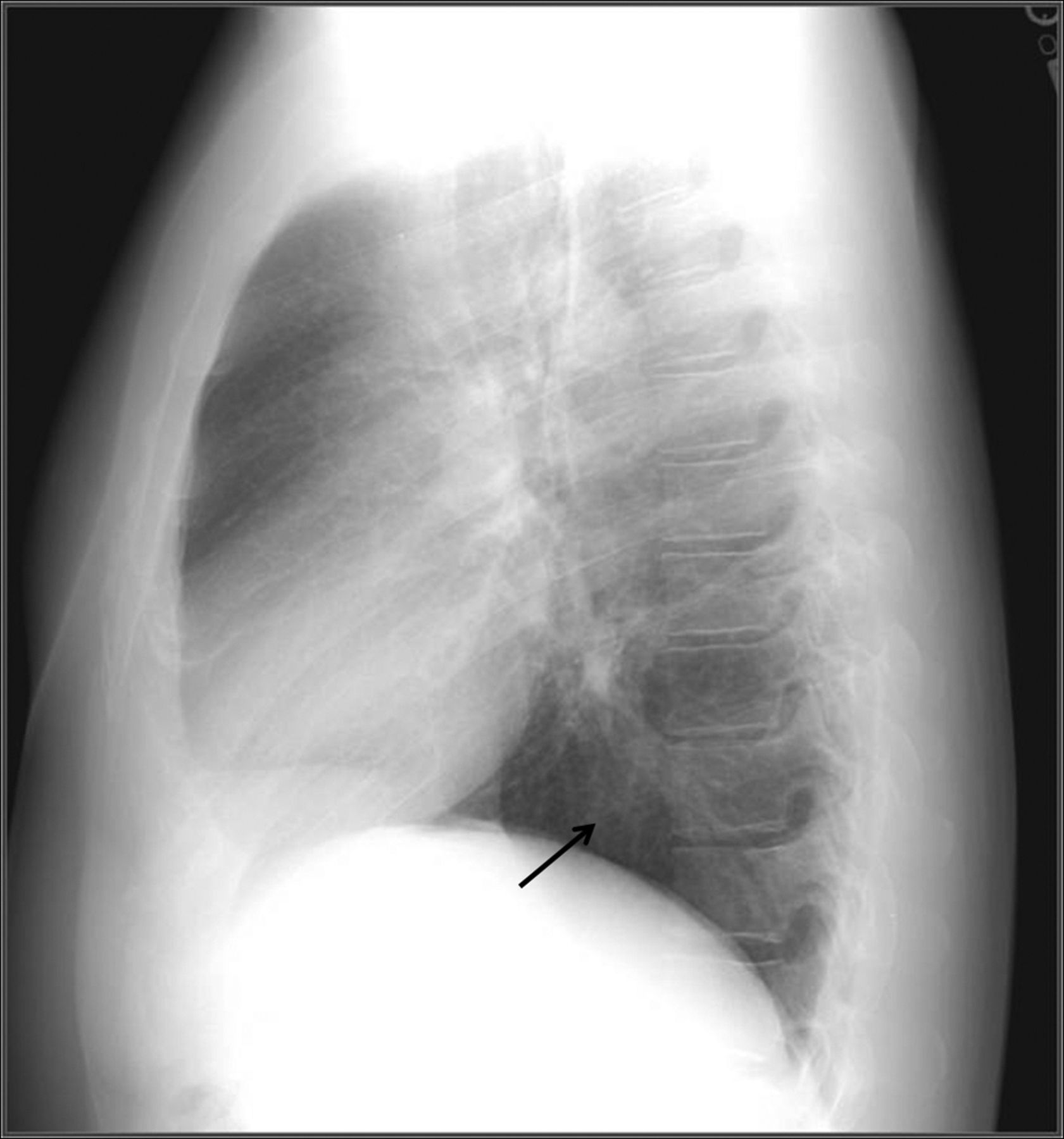
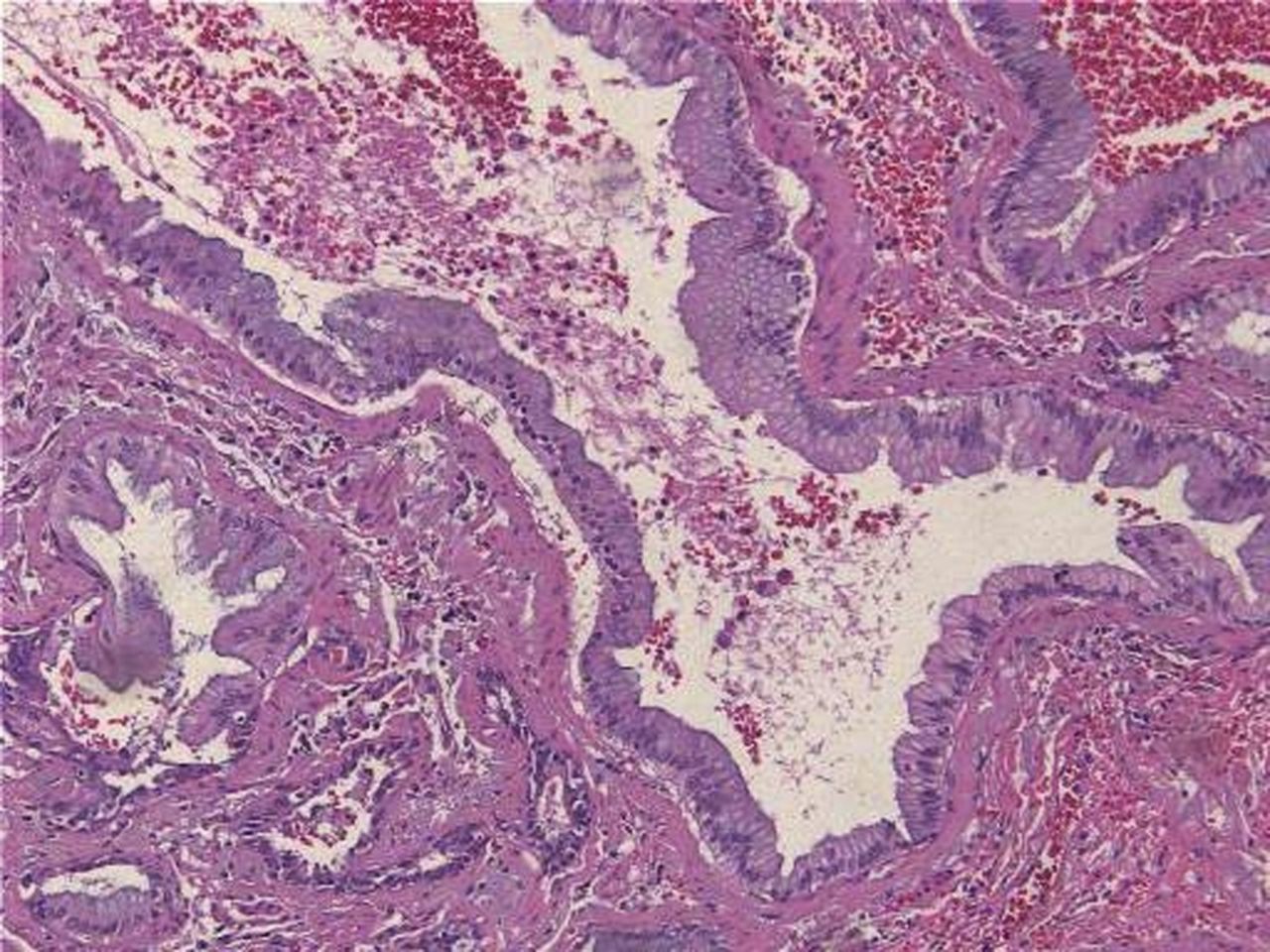
A 38-year-old white male presented with 4 days of acute onset, low volume, hemoptysis. He denied infectious, constitutional, or other respiratory symptoms. Social history was notable for a 50 pack-year smoking history. Physical examination was normal. Chest radiograph revealed an ill defined right lower lobe infiltrate (Figs. 1 and 2). Computed tomography of the chest demonstrated an irregular, thin walled, cystic lesion with adjacent nodularity and minimal calcification (Fig. 3). Bronchoscopy revealed airway distortion, without endobronchial abnormality, preventing examination of the right lower lobe airways below the superior segment take-off. Bronchial washings for cytology and bacterial, fungal, and mycobacterial cultures were negative. Due to his persistent hemoptysis and concerns for possible future infectious complications, the patient underwent a thoracotomy and right lower lobectomy (Fig. 4). Intraoperative course was complicated by multiple pleural adhesions, requiring extensive dissection and placement of surgicel to control bleeding at the costovertebral junction. The patient's postoperative course was complicated by back pain and lower extremity neurologic findings, prompting emergent decompressive laminectomy and removal of surgicel expansion into the spinal canal, with subsequent cord compression. Pathologic specimen demonstrated a 10 cm, mostly thin walled cyst with features suggestive of a CCAM and areas of adenocarcinoma (mixed subtype with acinar and bronchioloalveolar patterns) (Fig. 5). Microscopic evaluation demonstrated foci of fibrosis lining the cavity, with pseudostratified columnar epithelium that transitions to adenomatous disorganized tissue consistent with a type 1 CCAM and associated adenocarcinoma (see Figs. 5 and 6). Immunohistological staining was positive for thyroid transcription factor 1 (TTF-1), consistent with the diagnosis of a primary lung adenocarcinoma.

Fig. 1.
  • Download figure
  • Open in new tab
  • Download powerpoint
Fig. 1.

Posterior/anterior chest x-ray demonstrating an ill defined reticular and interstitial opacity that overlies the right middle and lower lobes.

Fig. 2.
  • Download figure
  • Open in new tab
  • Download powerpoint
Fig. 2.

Lateral chest x-ray demonstrating reticular and interstitial opacity in the right lower lobe.

Fig. 3.
  • Download figure
  • Open in new tab
  • Download powerpoint
Fig. 3.

Computed tomography of the chest demonstrating an irregular, cystic lesion and right lower lobe with nodular characteristics in the superior, posterior aspects. There is adjacent atelectasis, scarring , nodules, and hyperinflation. Adjacent ground-glass may be due to recent hemoptysis or inflammation.

Fig. 4.
  • Download figure
  • Open in new tab
  • Download powerpoint
Fig. 4.

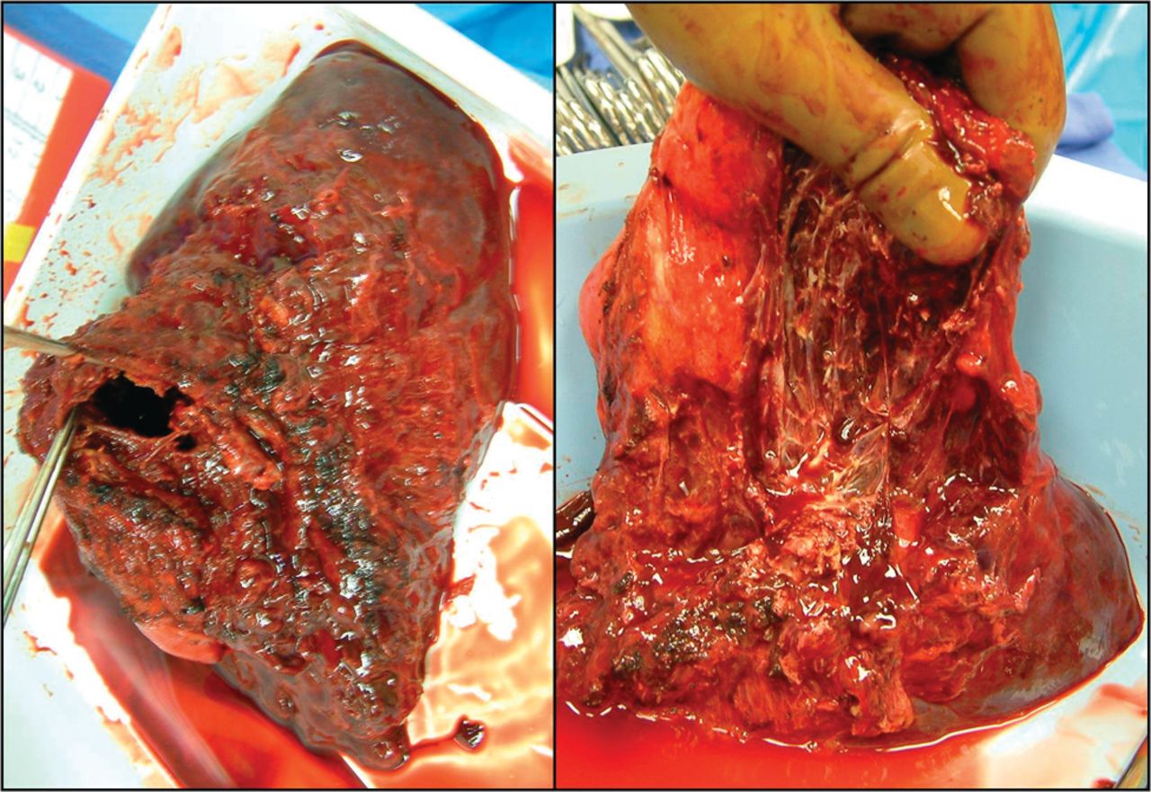
Pathologic specimen of the right lower lobe demonstrating a cavitary lesion with evidence of chronic pleural inflammation and adhesions.

Fig. 5.
  • Download figure
  • Open in new tab
  • Download powerpoint
Fig. 5.

Bronchoalveolar carcinoma lining normal alveolar structure.

Fig. 6.
  • Download figure
  • Open in new tab
  • Download powerpoint
Fig. 6.

Thick fibrous connective tissue with islands of cuboidal and pseudocolumnar epithelium.

Discussion

CCAMs were first identified in 1949 by Ch'in and Tang as distinct and rare lesions that occurred in stillborn or premature newborns with anasarca.2 CCAMs are the most common form of congenital parenchymal lung malformations and have an estimated incidence of 1:25,000–1:35,000 births.3 The differential diagnosis includes bronchogenic cyst, pulmonary sequestration, congenital diaphragmatic hernia, and congenital lobar emphysema. CCAMs were previously categorized according to the Stocker classifications 1, 2, and 3.4 More recently this classification was expanded and the name changed to congenital pulmonary airway malformation (CPAM), reflecting the fact that not all of these lesions are cystic (Table 1).1 The staging of CPAMs from type 0 to type 4 malformations reflects the progression of developmental abnormalities through the tracheobronchial tree, from large airways to bronchioles and alveoli.

View this table:
  • View inline
  • View popup
  • Download powerpoint
Table 1.

Expanded Stocker Classification for Congenital Pulmonary Airway Malformations

The pathophysiology of CPAM development is controversial. Some authors believe they result from arrested development of localized portions of the bronchial tree during the sixth and seventh week of fetal development. Others believe they are hamartomatous lesions of the bronchial tree.5,6 TTF1 is expressed solely in lung, thyroid, and certain portions of the brain. It regulates lung epithelial differentiation and plays a central role in lung development, and is used in immunohistochemical staining of malignant lesions to help differentiate primary lung cancer from metastatic.7 TTF-1 is found in normal lung tissue, but expression is solely found in bronchial and alveolar epithelial cells.8 Disruption of TTF-1 by either mutation or deletion has also been postulated to contribute to the development of CPAMs.9

CPAMs are usually identified during routine obstetric care due to the increased modern use of ultrasound, and children with this abnormality most commonly present with respiratory distress and recurrent pulmonary infections.10 The fetal lesions can remain stable in size, enlarge, or be associated with polyhydramnios or hydrops fetalis, which carries with it a poor prognosis.11,12 CPAMs account for 42% of pediatric lung resections, with 86% identified by age 5.1,7 To date, there have been 42 reported cases (Table 2) of CPAM presenting in adulthood (age > 17 y). In these cases there was near equal distribution between males (no. = 21) and females (no. = 21).12–24 The mean age on presentation was approximately 36 years old (range 17–65 years old). Adult CPAMs are most common in the lower lobe (no. = 30), with slight right (no. = 22) versus left (no. = 18) predominance. The most common presenting symptom in adults is recurrent pulmonary infections (no. = 21). Contrary to recent case reports on this subject, our review of the literature identified asymptomatic radiographic abnormalities as the second most common presentation (no. = 10, 24%) of CPAM in adults.14 Other complications include pneumothorax (no. = 5), hemoptysis (no. = 3), fever (no. = 1), dyspnea (no. = 1), and malignant transformation.3 The exact incidence of malignant transformation with CPAMs is unknown but suggested to be approximately 1%.3 A large case series in a pediatric population found a 4–9% association between CPAMs and lung tumors.25

View this table:
  • View inline
  • View popup
  • Download powerpoint
Table 2.

Reported Cases of Congenital Pulmonary Airway Malformation Presenting in Adulthood

Type 1 CPAMs have mucogenic epithelium, which is hypothesized likely to be the cause of neoplastic transformation.26 Bronchoalveolar cell carcinoma (BAC) is the most common form of malignant cell type associated with CPAM in adults, which can be challenging to identify using current available imaging modalities and non-surgical biopsy sampling.25 The vast majority of authors therefore recommend surgical excision of these lesions for definitive treatment, due to the risk of malignant transformation and recurrent pulmonary infection, the latter of which typically responds poorly to medical therapy. This recommendation remains controversial, as some authors argue that the risk of malignant transformation is overstated.25 There have been no systematic reviews published comparing conservative management to surgical resection, and, given the high incidence of asymptomatic presentation that our review has identified, there may be a tendency to consider serial follow-up only in these individuals. It is worth noting one published case report that identified a 6-year-old male with a type 1 CPAM and biopsy proven BAC. He was managed conservatively over a 15 year period and at age 21 he was diagnosed with metastatic adenocarcinoma.27

Although the number of adult cases of CPAMs and associated lung cancer are small, most of the literature to date support that there is a relationship between the two. Surgical resection provides definitive management of this rare condition and its high rate of complications, and carries with it a low risk in this otherwise healthy population. Despite the complicated postoperative course in this patient, surgery remains the logical treatment strategy of choice in these authors' opinion. The complication witnessed in this case is rare but reported. Given the tendency for recurrent, chronic infection and inflammation found in association with CPAMs, a potential complicated intraoperative dissection should be anticipated as part of preoperative planning. Given the risk of continued expansion found in our case, surgicel should not be used in and around the costovertebral junction.

Footnotes

  • Correspondence: Ryan McDonough DO CPT MC USA, Department of Medicine, Madigan Army Medical Center, Building 9040, Fitzsimmons Drive, Tacoma WA 98335. E-mail: ryan.j.mcdonough{at}us.army.mil.
  • The views expressed in this paper are those of the authors and do not reflect the official policy or position of the Department of the Army, Department of Defense, or the United States Government.

  • The authors have disclosed no conflicts of interest.

  • © 2012 by Daedalus Enterprises Inc.

References

  1. 1.↵
    1. Hammer SP
    1. Stocker JT
    . Congenital and developmental diseases in pulmonary pathology. In: Hammer SP editor. Pulmonary pathology. New York: Springer-Verlag; 1994:155–190.
  2. 2.↵
    1. Ch'in KT,
    2. Tang MY
    . Congenital adenomatoid malformation of one lobe of lung with general anasarca. Arch Pathol Lab Med 1949;48(3):221–229.
    OpenUrl
  3. 3.↵
    1. Lantuejoul S,
    2. Nicholson AG,
    3. Sartori G,
    4. Piolat C,
    5. Danel C,
    6. Brabencova E,
    7. et al
    . Mucinous cells in type 1 pulmonary cystic adenomatoid malformations as mucinous bronchioloalveolar carcinoma precursors. Am J Surg Pathol 2007;31(6):961–969.
    OpenUrlCrossRefPubMed
  4. 4.↵
    1. Taussig LM,
    2. Landau LI
    1. Clements BS
    . Congenital malformations of the lungs and airways. In: Taussig LM, Landau LI editors. Pediatric respiratory medicine. St. Louis: Mosby; 1999:1106–1037.
  5. 5.↵
    1. Stocker JT,
    2. Dehner LP
    1. Stocker JT
    . The respiratory tract. In Stocker JT, Dehner LP editors. Pediatric pathology , 2nd edition. Philidelphia: Lippincott, Williams & Wilkins; 2001:445–517.
  6. 6.↵
    1. Whitsett JA
    . Genetic disorders influencing lung formation and function at birth. Hum Mol Genet 2004;13(2):R207–R215.
    OpenUrlAbstract/FREE Full Text
  7. 7.↵
    1. Ordóñez NG
    . Thyroid transcription factor-1 is a marker of lung and thyroid carcinomas. Adv Anat Pathol 2000;7(2):123–127.
    OpenUrlPubMed
  8. 8.↵
    1. Fabbro D,
    2. Di Loreto C,
    3. Stamerra O,
    4. Alberto Beltrami C,
    5. Lonigro R
    . Damante G. TTF-1 gene expression in human lung tumours. Eur J Cancer 1996;32(6):512–517.
    OpenUrlCrossRef
  9. 9.↵
    1. Shanmugan G,
    2. MacArthur K,
    3. Pollock JCS
    . Congenital lung malformations-antenatal and postnatal evaluation and management. Eur J Cardiothorac Surg 2005;27(1):45–52.
    OpenUrlAbstract/FREE Full Text
  10. 10.↵
    1. Laberge JM,
    2. Bratu I,
    3. Flageole H
    . The management of asymptomatic congenital lung malformations. Paediatr Respir Rev 2005;5(Suppl A):S305–S312.
    OpenUrl
  11. 11.↵
    1. Hancock BJ,
    2. Di Lorenzo M,
    3. Youssef S,
    4. Yazbeck S,
    5. Marcotte JE,
    6. Collin P
    . Childhood primary neoplasms. J Pediatr Surg 1993;28(9):1133–1136.
    OpenUrlCrossRefPubMed
  12. 12.↵
    1. Yamashiro T,
    2. Murayama S,
    3. Nakayama T,
    4. Akamine T,
    5. Moromizato H,
    6. Hirayasu,
    7. Yara S
    . Congenital cystic adenomatoid malformation presenting as repeated pneumonia in a young adult. Radiat Med 2007;25(9):488–491.
    OpenUrlPubMed
  13. 13.
    1. Oh B,
    2. Lee J,
    3. Kim J,
    4. Lim C,
    5. Koh Y
    . Congenital cystic adenomatoid malformation of the lung in adults: clinical and CT evaluation of seven patients. Respirology 2006;11(4):496–501.
    OpenUrlCrossRefPubMed
  14. 14.↵
    1. Lai P,
    2. Cohen D,
    3. DeCamp M,
    4. Fazio S,
    5. Roberts D
    . A 40-year-old woman with an asymptomatic cystic lesion in her right lung. Chest 2009;136(2):622–627.
    OpenUrlCrossRefPubMed
  15. 15.
    1. Vicidomini G,
    2. Santini M,
    3. Baldi A,
    4. Cesarano T,
    5. Di Marino MP,
    6. Baldi F
    . Cystic adenomatoid malformation of the lung in an adult. Minerva Chir 1997;52(4):469–473.
    OpenUrlPubMed
  16. 16.
    1. Herrero Y,
    2. Pinilla I,
    3. Torres I,
    4. Nistal M,
    5. Pardo M,
    6. Gomez N
    . Cystic adenomatoid malformation of the lung presenting in adulthood. Ann Thorac Surg 2005;79(1):326–329.
    OpenUrlCrossRefPubMed
  17. 17.
    1. Davies A,
    2. Bapat V,
    3. Treasure T
    . Adult presentation of congenital cysticadenomatoid malformation: successful surgical management. J Thorac Cardiovasc Surg 2006;132(6):1493–1494.
    OpenUrlCrossRefPubMed
  18. 18.
    1. Khan N,
    2. Jones M,
    3. Greaves M
    . Congenital cystic adenomatoid malformation of an entire lung in a 33-year-old man: a case report and review of the literature. Brit J Radiol 2008;81(971):276–278.
    OpenUrl
  19. 19.
    1. Huang H,
    2. Talbot R,
    3. Liu K,
    4. Chen C,
    5. Fang H
    . Infected cystic adenomatoid malformation in an adult. Ann Thorac Surg 2004;78(1):337–339.
    OpenUrlCrossRefPubMed
  20. 20.
    1. Morelli L,
    2. Piscioli I,
    3. Licci S,
    4. Donato S,
    5. Catalucci A,
    6. Del Nonno F
    . Pulmonary congenital cystic adenomatoid malformation, type I, presenting as a single cyst of the middle lobe in an adult: case report. Diagn Pathol 2007;2:17.
    OpenUrlCrossRefPubMed
  21. 21.
    1. Lujan M,
    2. Bosque M,
    3. Mirapeix R,
    4. Marco T,
    5. Asensio O,
    6. Domingo C
    . Late-onset congenital cystic adenomatoid malformation of the lung. Respiration 2002;69(2):148–154.
    OpenUrlCrossRefPubMed
  22. 22.
    1. Hellmuth D,
    2. Glerant JC,
    3. Sevestre H,
    4. Remond A,
    5. Jounieaux V
    . Pulmonary adenomatoid malformation presenting as unilobar cysts in an adult. Respir Med 1998;92(12):1364–1367.
    OpenUrlCrossRefPubMed
  23. 23.
    1. Dahabreh J,
    2. Zisis C,
    3. Arnogiannaki N
    . Congenital cystic adenomatoid malformation in an adult presenting as lung abscess. Eur J Cardiothorac Surg 2000;18(6):720–723.
    OpenUrlAbstract/FREE Full Text
  24. 24.↵
    1. West D,
    2. Nicholson AG,
    3. Colquhoun I,
    4. Pollock J
    . Bronchioloalveolar carcinoma in congenital cystic adenomatoid malformation of lung. Annal Thorac Surg 2007;83(2):687–689.
    OpenUrlCrossRefPubMed
  25. 25.↵
    1. Fitzgerald DA
    . Congenital cyst adenomatoid malformations: resect some and observe all? Paediatr Respir Rev 2007;8(1):67–76.
    OpenUrlCrossRefPubMed
  26. 26.↵
    1. Morresi A,
    2. Wockel W,
    3. Kark O
    . Adenomatoid cystic lung abnormality in adults with associated bronchoalveolar carcinoma. Pathologe 1995;16(4):292–298.
    OpenUrlCrossRefPubMed
  27. 27.↵
    1. Loachimescu OC,
    2. Mehta AC
    : From cystic pulmonary airway malformation, to bronchioloalveolar carcinoma and adenocarcinoma of the lung. Eur Respir J 2005;26(6):1181–1187.
    OpenUrlAbstract/FREE Full Text
PreviousNext
Back to top

In this issue

Respiratory Care: 57 (2)
Respiratory Care
Vol. 57, Issue 2
1 Feb 2012
  • Table of Contents
  • Table of Contents (PDF)
  • Cover (PDF)
  • Index by author


Print
Download PDF
Article Alerts
Sign In to Email Alerts with your Email Address
Email Article

Thank you for your interest in spreading the word on American Association for Respiratory Care.

NOTE: We only request your email address so that the person you are recommending the page to knows that you wanted them to see it, and that it is not junk mail. We do not capture any email address.

Enter multiple addresses on separate lines or separate them with commas.
Congenital Pulmonary Airway Malformation: A Case Report and Review of the Literature
(Your Name) has sent you a message from American Association for Respiratory Care
(Your Name) thought you would like to see the American Association for Respiratory Care web site.
CAPTCHA
This question is for testing whether or not you are a human visitor and to prevent automated spam submissions.
Citation Tools
Congenital Pulmonary Airway Malformation: A Case Report and Review of the Literature
Ryan J McDonough, Alexander S Niven, Keith A Havenstrite
Respiratory Care Feb 2012, 57 (2) 302-306; DOI: 10.4187/respcare.00727

Citation Manager Formats

  • BibTeX
  • Bookends
  • EasyBib
  • EndNote (tagged)
  • EndNote 8 (xml)
  • Medlars
  • Mendeley
  • Papers
  • RefWorks Tagged
  • Ref Manager
  • RIS
  • Zotero

Share
Congenital Pulmonary Airway Malformation: A Case Report and Review of the Literature
Ryan J McDonough, Alexander S Niven, Keith A Havenstrite
Respiratory Care Feb 2012, 57 (2) 302-306; DOI: 10.4187/respcare.00727
del.icio.us logo Digg logo Reddit logo Twitter logo CiteULike logo Facebook logo Google logo Mendeley logo
  • Tweet Widget
  • Facebook Like
  • Google Plus One

Jump to section

  • Article
    • Abstract
    • Introduction
    • Case Report
    • Discussion
    • Footnotes
    • References
  • Figures & Data
  • Info & Metrics
  • References
  • PDF

Related Articles

Cited By...

Keywords

  • congenital cystic adenomatoid malformation
  • CCAM
  • congenital lobar hyperinflation
  • bronchogenic cyst
  • lobar sequestration
  • hemoptysis

Info For

  • Subscribers
  • Institutions
  • Advertisers

About Us

  • About the Journal
  • Editorial Board

AARC

  • Membership
  • Meetings
  • Clinical Practice Guidelines

More

  • Contact Us
  • RSS
American Association for Respiratory Care

Print ISSN: 0020-1324        Online ISSN: 1943-3654

© Daedalus Enterprises, Inc.

Powered by HighWire