

Introduction
Primary tracheobronchial tumors are rare neoplasms. They frequently develop in the trachea, carina, and endobronchial regions.1 Because they are rare and because of the variation in their histogenesis, chest x-ray findings and clinical and operational outcomes have not been well elucidated.2,3 Endobronchial tumors may be benign or show low- or high-grade malignancy.4–6 Affected patients generally present with symptoms of either asthma or infection that has developed as a result of obstruction.2,4,6 However, these symptoms are nonspecific. We herein report 4 such affected subjects in our clinic and discuss their symptoms, pathological findings, and treatments.
Case Summary
Case 1
A 31-y-old female subject had been treated for asthma for 5 y. Although she was using high doses of inhaled steroids, β mimetics, montelukast, and theophylline, her symptoms continued. Physical examination revealed decreased breath sounds on the left side. Thoracic computed tomography (CT) was performed to follow up the observed radiolucency in the left lung on her chest x-ray. Endobronchial narrowing of the left main bronchus and resultant air trapping were found on thoracic CT. Rigid bronchoscopy revealed a lesion completely occluding the left main bronchus. A biopsy was performed, and the pathological findings revealed an adenoid cystic carcinoma (Fig. 1). She was treated with open surgery. Her symptoms and physical examination findings improved after surgery. We discontinued asthma treatment.

Case 1: histopathological image of adenoid cystic carcinoma of the lung (hematoxylin and eosin stain, magnification × 100).
Case 2
A 23-y-old male subject with a 5-pack-year cigarette history had been treated for asthma for 4 y. He was using asthma medications irregularly. Physical examination revealed bilateral rhonchi. His chest x-ray was normal. Thorax CT revealed an endobronchial lesion in the right main bronchus, and follow-up bronchoscopy revealed a pedunculated, hemorrhage-prone mass lesion almost completely occluding the right main bronchus. A biopsy revealed a typical carcinoid tumor (Figs. 2 and 3). He was treated with interventional bronchoscopy with electrocautery.

Case 2: mass lesion almost entirely blocking the right main bronchus.
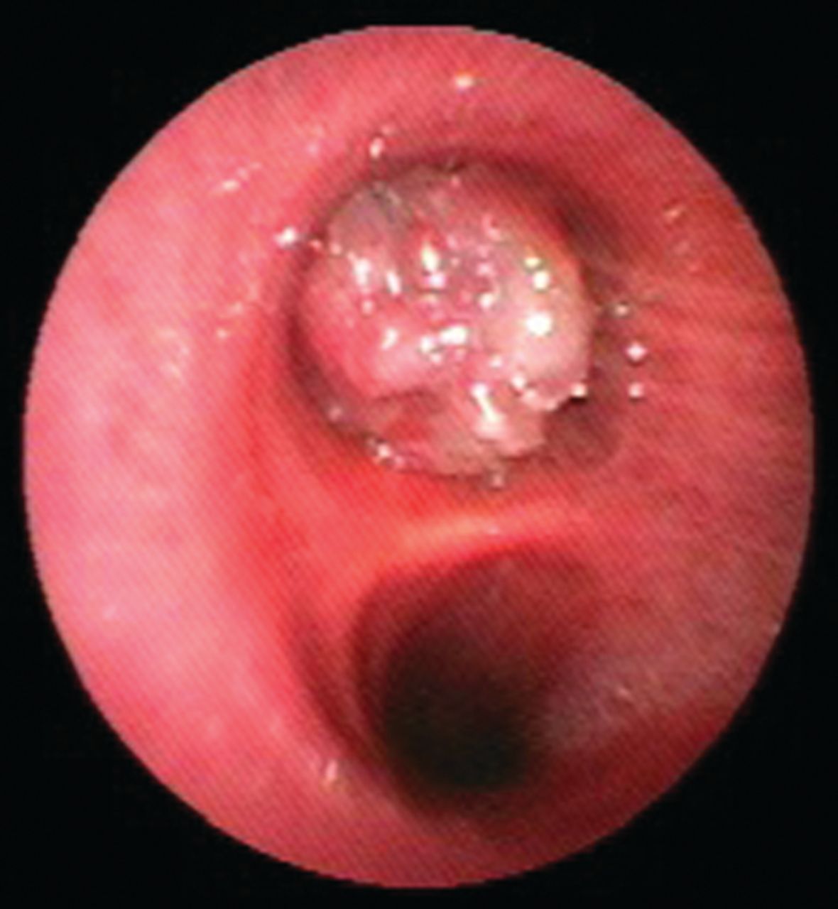
Case 2: bronchoscopic finding of pedunculated mass lesion almost entirely blocking the right main bronchus.
Case 3
A 55-y-old male subject had been treated for asthma for 2 y. Although he was using high doses of inhaled steroids, β mimetics, and montelukast, his symptoms continued. Physical examination revealed decreased breath sounds on the right side. Chest x-ray revealed right hilar enlargement, and thoracic CT showed an ∼2-cm lesion in his right main bronchus. Bronchoscopy showed two mass lesions almost completely occluding the right main bronchus. The biopsy result revealed adenoid cystic carcinoma (Fig. 4). He was treated with open surgery.

Case 3: 2 mass lesions blocking the right main bronchus.
Case 4
A 30-y-old female subject had been treated for asthma for 4 y. Her symptoms continued despite her regular use of asthma medications. Her chest x-ray was normal. Thoracic CT revealed an endobronchial lesion in the right main bronchus that did not entirely occlude the lumen. The pathological result was adenoid cystic carcinoma. She was treated with open surgery.
Discussion
Clinically, endobronchial tumors cause air trapping or secondary lung infections.2–6 Their diagnosis is usually delayed because they do not appear on chest x-rays. CT and bronchoscopy are helpful in obtaining a diagnosis in these cases.4 In affected patients, interventional bronchoscopy is critical for timely treatment and diagnosis. All of our subjects were being treated for asthma but did not respond adequately to treatment. None of our subjects presented with secondary infection.
Tracheobronchial tumors may be benign or show low- or high-grade malignancy. Therefore, CT is useful in revealing lesions not expected to be associated with these tumors. Solitary papillomas, mucous tissue adenomas, inflammatory myofibroblastic tumors, schwannomas, leiomyomas, hamartomas, hemangiomas, and chondromas from benign tumors have been reported.3,4,7–10
Squamous cell cancer of the tracheobronchial tree, carcinoid tumors, adenoid cystic carcinomas, and mucoepidermoid carcinomas from low-grade malignant tumors have also been reported.2–4,6,11,12 These tumors are low grade but can recur after treatment or can result in distant metastasis. Three of our subjects had adenoid cystic carcinomas, and one had a carcinoid tumor. CT revealed no lymph node metastasis or distant metastasis in any subject. Because our subjects were identified in 2012 and 2013, we believe that it is too early to evaluate them for recurrence; however, recurrence has not been observed in the 4 subjects to date.
Most of the 14 subjects with endobronchial and adenoid cystic carcinomas examined by Albers et al13 had nonspecific breathing complaints such as cough and dyspnea due to narrowing of the airways; however, few subjects had asthma anamnesis with long-term wheezing and stridor complaints. Nine of these subjects were female, and 5 were male. Of our subjects, 2 were female, and 2 were male.
Dewan et al14 reported that the right side was most frequently affected in 31 subjects with tracheobronchial carcinoid tumors. The tumor was present in a main bronchus in 30 of these 31 subjects; one patient had a tracheal tumor. Most of these subjects had typical carcinoid tumors. In our subject with a carcinoid tumor, the tumor was located at the entrance of the right main bronchus, and its pathology was consistent with a typical carcinoid tumor.
Moran et al15 reported that 11 of 16 subjects with adenoid cystic carcinoma were male, and 5 were female. Of these subjects, 7 underwent pneumonectomy, 6 underwent lobectomy, and 2 underwent lobectomy and chemotherapy. One subject underwent only chemotherapy because of disease progression. For our subjects, mass excision only during open surgery was performed in cases 1–3 with no resection. Case 2 also underwent interventional bronchoscopy with electrocautery. The symptoms and physical examination findings of all of our subjects improved after intervention.
In conclusion, tracheobronchial tumors, endobronchial adenoid cystic carcinomas, and carcinoid tumors should be suspected in patients with chronic coughing or frequent lung infections who undergo long-term treatment for asthma and who do not respond well to asthma medications. Even if the chest x-ray findings are normal, thoracic CT and, if necessary, bronchoscopy should be performed.
Teaching Points
Chest CT, as well as bronchoscopy, should be performed in treatment-resistant patients with asthma, even in those with a normal chest x-ray.
Endobronchial tumors should be suspected in patients with chronic coughing or frequent lung infections who undergo long-term treatment for asthma.
Footnotes
- Correspondence: Ahmet Arısoy, Pulmonary Medicine Department, Private Istanbul Hospital, 65100 Istanbul, Turkey. E-mail: drahmetarisoy{at}gmail.com.
The authors have disclosed no conflicts of interest.
- Copyright © 2014 by Daedalus Enterprises


