Article Text

Abstract
Non-invasive respiratory support is increasingly popular but is associated with complications including nasal trauma. The present report describes a novel method of oral continuous positive airway pressure (CPAP) delivery in an extremely premature infant with severe nasal septum erosion.
The distal end of a cut down endotracheal tube was passed through a small hole made in the teat of a dummy (infant pacifier) and sutured in place. The dummy was secured in the infant's mouth and CPAP was delivered to the pharynx. The device was well tolerated and the infant was successfully managed using this technique for 48 days, avoiding endotracheal intubation and ventilation.
Statistics from Altmetric.com
Case report
A girl infant born at 26 weeks' gestation and weighing 575 g developed severe respiratory distress syndrome requiring treatment with conventional ventilation and high-frequency oscillatory ventilation for 13 days.
On day 13 the infant was extubated and managed with non-synchronised nasal intermittent positive pressure ventilation (NIPPV), using a Drager Babylog 8000 plus ventilator (Drager Medical, Lubeck, Germany), via binasal Hudson prongs, size 0 (Teleflex Medical, Research Triangle Park, North Carolina, USA). Maximal NIPPV settings were a peak inflation pressure of 20 cm H2O, end expiratory pressure of 9 cm H2O, inflation rate of 30/min and inflation time of 0.3 s. Oxygen requirements rose to 60% 9 days following extubation. In order to avoid reintubation, the infant was treated with a 10-day course of low-dose dexamethasone.1 She responded and remained stable on NIPPV with an oxygen requirement of 25% to 35%. On the 12th day of non-invasive ventilation, the clinical team noted significant breakdown of the skin on the nasal septum and a trial of nasal mask (Infant Flow System Small Mask, Cardinal Health, Dublin, Ohio, USA) NIPPV was given. Mask NIPPV was continued for 10 days, but resulted in ongoing trauma to the area of skin breakdown and in particular deterioration in the condition of the nasal septum (figure 1A).

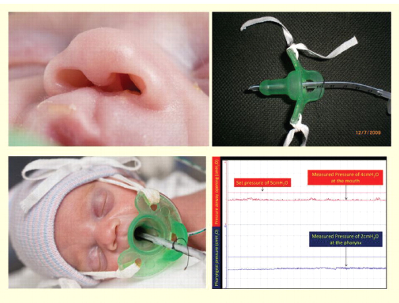
A. Premature infant with nasal septum erosion. B. Dummy continuous positive airway pressure (CPAP) device. C. Oral CPAP in place on the infant. D Pharyngeal pressure measurements.
On day 36, as an alternative to intubation and ventilation, it was decided to try a form of oral continuous positive airway pressure (CPAP). It was hoped this would allow time for wound healing while continuing non-invasive positive pressure respiratory support. A preterm infant pacifier (dummy) (Gumdrop Preemie Pacifier, Hawaii Medical, Pembroke, Massachusetts, USA) was modified by cutting a small hole in the tip of the teat. A 3-mm diameter endotracheal tube (ETT) was passed through the hole in the teat so that the tip and side port of the ETT protruded just beyond the teat (figure 1B). The ETT was stitched to the side of the dummy at 5 cm to prevent advancement of the ETT through the hole in the teat. The dummy was placed in the infant's mouth and secured in position with ties attached to the infant's bonnet (figure 1C). The tip of the ETT was estimated to be in the oropharynx just beyond the hard palate.
What is already known on this topic
Nasal continuous positive airway pressure (CPAP) is frequently used in extremely low birthweight infants.
Nasal trauma is a complication of nasal CPAP.
What this study adds
Oral CPAP is a new but an unvalidated interface device that can provide ongoing CPAP in the setting of nasal trauma.
The NIPPV settings continued unchanged, the infant remained clinically stable and there was no change in oxygen requirement following commencement of oral CPAP. Apnoeas and bradycardias were reported before and after the change to oral CPAP and there was no noticeable change in their frequency. The NIPPV inflation rate was gradually reduced to 10 per min, and then stopped, to provide CPAP alone. The oral tube was used to deliver first NIPPV and then CPAP for a total of 48 days. During this time the infant was fed via an orogastric tube passed alongside the dummy. The delivery system required intermittent suctioning and had to be replaced if there was vomiting to prevent milk from blocking the ETT. The device was well tolerated and no major complications were observed.
To assess the effectiveness of this method of CPAP delivery, simultaneous pharyngeal and CPAP circuit pressures were measured, using calibrated pressure sensors. This was undertaken just prior to discontinuation of CPAP, when the set pressure was 5 cm H2O. The CPAP circuit pressure measured 4 cm H2O and pressures of 2 cm H2O were recorded using a feeding tube placed in the pharynx (figure 1D).
Positive pressure support was discontinued after 71 days (combination of NIPPV and CPAP), by which time there was good healing of the nasal wound (figure 1A). The infant was discharged home at 5 weeks post term, with home oxygen, with a plan for later surgical reconstruction of the nasal septum.
Discussion
Non-invasive respiratory support is being used earlier and more frequently for the treatment of respiratory disease in preterm infants. Consequently very low and extremely low birthweight infants may be on NIPPV and CPAP for long periods of time. It is therefore possible that more complications related to nasal CPAP use will be seen and alternate methods of CPAP delivery may need to be explored.
When using prongs to deliver CPAP we know that short binasal prongs are the most effective, although the optimal CPAP delivery device is yet to be established.2 We do not know the optimal delivery device for NIPPV, nor are we sure of the efficacy of nasal masks for CPAP delivery in premature infants.
Nasal trauma is a well documented complication of non-invasive respiratory support3,–,5 and its incidence has been reported as high as 40%.4 In 2003 Yong et al3 compared the Infant Flow Driver (IFD, Cardinal Health, Dublin, Ohio, USA) silicon nasal mask with the IFD nasal prongs in 89 infants. No significant difference in the incidence of trauma was seen (29% in masks vs 35% with prongs, p=0.5), although different sites of injury were observed. Nasal prong trauma was seen most commonly on the medial aspect of the nostrils at the septum, whereas nasal mask trauma was seen most often at the base of the nasal septum, near the philtrum. In addition to crusting, bleeding, redness and excoriation at these sites, both devices also caused narrowing of the nasal passages. Yong reported that rates of trauma increased with length of use of CPAP, whatever the mode of its delivery. However injury to the columella during short binasal prong CPAP has been reported as early as 3 days after commencement of CPAP.5
Significant nasal injury may limit the use of nasal CPAP in some preterm infants who need ongoing respiratory support and an alternative delivery method may be of benefit. Oral CPAP has not been previously described in preterm infants although in the early 1970s face masks were strapped to the infant to provide CPAP. Recently an oral CPAP mask, for use in obstructive sleep apnoea, has been described in adults.6 In adults, similar pressures were achieved with the oral mask compared with established nasal routes.6
We measured a pressure drop of 2 cm H2O from the CPAP circuit to the pharynx, which is similar to the pressure difference described by De Paoli et al during nasal prong CPAP,7 providing encouragement that oral CPAP could be as effective as nasal CPAP. In our case oral CPAP appeared clinically effective and was well tolerated, it was manageable over a prolonged period of time and there was no evidence of harm. The use of oral CPAP avoided the need for intubation and ventilation, with its inherent complications, while allowing healing of the nasal mucosa.
When nasal trauma occurs in premature infants who are CPAP dependent, alternative strategies include using a single nasopharyngeal prong as an interface or soft binasal prongs connected to a high flow system. In our case, a single prong would have interfered with the healing of the nasal septum and potentially aggravated the injury, as we had observed using the nasal mask. High flow systems are increasingly being used in neonatal intensive care units and although potentially useful in the setting of nasal trauma, currently available systems at present do not measure or regulate the delivered pressure or provide NIPPV.
In summary, we report a new method of CPAP delivery in a preterm infant with nasal injury without adverse effects. This strategy allowed healing of the nasal septum while successfully avoiding reintubation of the infant. We propose that this could be an alternative method of providing non-invasive positive pressure ventilation, and may be useful for those infants where nasal CPAP with prongs or mask is not possible. However, oral CPAP is a new device that requires additional evaluation. Further research is required to define the safety and efficacy of this technique before it is more widely used for cases of nasal trauma.
Footnotes
-
Competing interests None.
-
Provenance and peer review Not commissioned; externally peer reviewed.
-
Patient consent Obtained.