 

Main menu

  • Home
  • Content
    • Current Issue
    • Editor's Commentary
    • Archives
    • Most-Read Papers of 2022
  • Authors
    • Author Guidelines
    • Submit a Manuscript
  • Reviewers
    • Reviewer Information
    • Create Reviewer Account
    • Reviewer Guidelines: Original Research
    • Reviewer Guidelines: Reviews
    • Appreciation of Reviewers
  • CRCE
    • Through the Journal
    • JournalCasts
    • AARC University
    • PowerPoint Template
  • Open Forum
    • 2023 Call for Abstracts
    • 2022 Abstracts
    • Previous Open Forums
  • Podcast
    • English
    • Español
    • Portugûes
    • 国语
  • Videos
    • Video Abstracts
    • Author Interviews
    • Highlighted Articles
    • The Journal

User menu

  • Subscribe
  • My alerts
  • Log in

Search

  • Advanced search
American Association for Respiratory Care
  • Subscribe
  • My alerts
  • Log in
American Association for Respiratory Care

Advanced Search

  • Home
  • Content
    • Current Issue
    • Editor's Commentary
    • Archives
    • Most-Read Papers of 2022
  • Authors
    • Author Guidelines
    • Submit a Manuscript
  • Reviewers
    • Reviewer Information
    • Create Reviewer Account
    • Reviewer Guidelines: Original Research
    • Reviewer Guidelines: Reviews
    • Appreciation of Reviewers
  • CRCE
    • Through the Journal
    • JournalCasts
    • AARC University
    • PowerPoint Template
  • Open Forum
    • 2023 Call for Abstracts
    • 2022 Abstracts
    • Previous Open Forums
  • Podcast
    • English
    • Español
    • Portugûes
    • 国语
  • Videos
    • Video Abstracts
    • Author Interviews
    • Highlighted Articles
    • The Journal
  • Twitter
  • Facebook
  • YouTube
Case ReportCase Reports

Hidden Diagnosis of Tuberculous Pleurisy Masked by Concomitant Pseudomonas oryzihabitans Bacteremia

Te-Yu Lin, Shih-Wei Wu, Gen-Min Lin and Yu-Guang Chen
Respiratory Care February 2012, 57 (2) 298-301; DOI: https://doi.org/10.4187/respcare.01103
Te-Yu Lin
Division of Infectious Diseases and Tropical Medicine;
  • Find this author on Google Scholar
  • Find this author on PubMed
  • Search for this author on this site
  • For correspondence: [email protected]
Shih-Wei Wu
Division of Pulmonary Medicine, Department of Internal Medicine, Tri-Service General Hospital, National Defense Medical Center, Taipei, Taiwan.
  • Find this author on Google Scholar
  • Find this author on PubMed
  • Search for this author on this site
Gen-Min Lin
Department of Internal Medicine, Hualien Armed Forces General Hospital, Hualien, Taiwan.
  • Find this author on Google Scholar
  • Find this author on PubMed
  • Search for this author on this site
Yu-Guang Chen
Division of Infectious Diseases and Tropical Medicine;
  • Find this author on Google Scholar
  • Find this author on PubMed
  • Search for this author on this site
  • Article
  • Figures & Data
  • References
  • Info & Metrics
  • PDF
Loading

Abstract

The clinical presentations of tuberculous pleurisy are usually nonspecific and have an insidious course, thus resulting in diagnostic challenges. Pseudomonas oryzihabitans is a nonfermenting, oxidase-negative, catalase-positive, Gram-negative bacillus that has rarely been encountered as a human pathogen. We present the case of a 30-year-old male patient who exhibited intermittent fever despite antibiotic treatment for Pseudomonas oryzihabitans bacteremia for 6 days. Tuberculous pleurisy was finally diagnosed by histopathologic and microbiologic studies. He recovered after a 2-week antibiotic course and 6-month antituberculosis treatment.

  • Pseudomonas oryzihabitans
  • bacteremia
  • tuberculous pleurisy

Introduction

Tuberculosis remains a major health problem worldwide. Extrapulmonary involvement is observed in less than 30% of all tuberculosis patients.1 Tuberculous pleurisy has an insidious course and nonspecific presentation, thus resulting in difficulties in diagnosis and subsequent treatment delays. Polymerase chain reaction of Mycobacterium tuberculosis (TB-PCR) is a rapid method for diagnosing pleural tuberculosis.2 However, if a patient is co-infected with other pathogens and the initial TB-PCR result is negative, diagnosis of pleural tuberculosis may be delayed. Here, we present the case of a patient with tuberculous pleurisy concomitant with Pseudomonas oryzihabitans bacteremia, who was successfully treated with antibiotics and antituberculosis treatment.

Case Report

A previously healthy 30-year-old man had experienced right pleuritic chest pain for one week, 2 months prior to this admission. He visited an emergency department and underwent a chest radiography examination (Fig. 1) that revealed increased lung markings in the right lower lung zone. Other laboratory parameters were within normal limits. Acute bronchitis was diagnosed by the emergency physician. His symptom was relieved by nonsteroidal anti-inflammatory drug treatment. He had high fever, chills, and right chest pain 2 days before admission to our hospital.

Fig. 1.
  • Download figure
  • Open in new tab
  • Download powerpoint
Fig. 1.

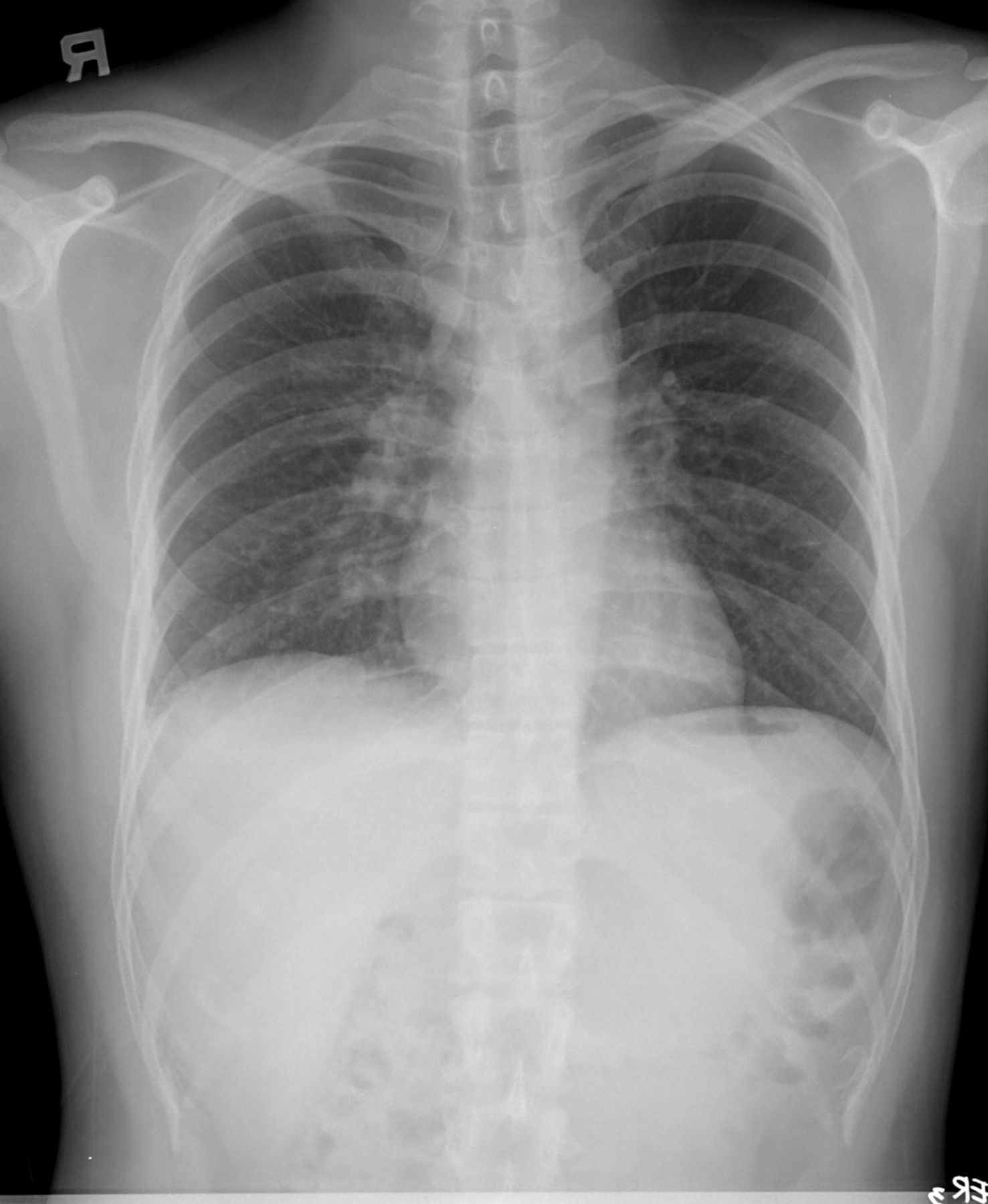
Chest radiograph showing increased lung markings in the right lower lung zone (obtained 2 months before admission).

On admission, his blood pressure was 112/77 mm Hg, pulse rate was 108 beats/min, respiration rate was 18 breaths/min, and body temperature was 38.6°C. A physical examination revealed decreased breathing sounds in the right lung field. Laboratory findings were as follows: white cell count 9.3 × 10³ cells/mL, proportion of neutrophils 80.5%, proportion of lymphocytes 10.1%, hemoglobin 15.9 g/dL, platelets 273 × 103 cells/mL, and C-reactive protein 6.85 mg/dL.

Chest radiography disclosed a right-sided pleural effusion (Fig. 2), and diagnostic thoracentesis was performed. Pleural effusion samples were obtained, and cell counting, biochemistry, pH evaluation, Gram staining, bacterial culture, acid-fast staining, M. tuberculosis culture (conventional Lowenstein Jensen medium), and TB-PCR were performed. The findings indicated lymphocyte predominance and revealed a yellow, turbid exudate. Empyema was suspected because of the low pH value, and a chest tube was inserted. Ceftazidime (2 g) was administered intravenously every 8 hours. Two days later, a Gram-negative bacillus was detected in the blood culture and finally identified as P. oryzihabitans (susceptible to amikacin, gentamicin, ciprofloxacin, ceftriaxone, ceftazidime, and imipenem). The patient exhibited intermittent fever under antibiotic treatment for 2 days, but the sputum and pleural fluid findings for bacteria, including M. tuberculosis (acid-fast stain and PCR), were all negative. A diagnostic procedure of video-assisted thoracoscopy with pleural biopsy was performed on the third day because of high suspicion of tuberculous pleural effusion. Multiple small white nodules scattered over the lung surface and parietal pleura regions were noted. Biopsy samples were sent for histopathologic examination, bacterial culture, and tuberculosis study. The pathological findings were suggestive of chronic granulomatous inflammation of the pleural tissue, with multinucleated giant cell formation, and TB-PCR of pleural tissue was still negative. Antituberculosis agents were given on the seventh day, based on pathological findings. The chest tube was removed on the tenth day, and the patient was discharged on the thirteenth day.

Fig. 2.
  • Download figure
  • Open in new tab
  • Download powerpoint
Fig. 2.

Chest radiograph showing right-side pleural effusion (obtained on the day of admission).

The patient was followed up in the out-patient department. Because P. oryzihabitans infection most commonly occurs in immunocompromised patients, we also examined the patient's immune status, including administration of a human immunodeficiency virus screen test and cell immune function tests, including tests of T lymphocyte subsets (CD3, CD4, CD8, and CD4/CD8 ratio). The human immunodeficiency virus screen test was negative, and the CD4/CD8 ratio was 0.64 (normal limit 1–2). Finally, 2 months after the patient was discharged, pleural effusion culture for M. tuberculosis was found to be positive. The patient recovered after a 2-week course of antibiotics and antituberculosis treatment (intensive phase: rifampin, pyrazinamide, isoniazid, and ethambutol; maintenance phase: rifampin, isoniazid, and ethambutol) for 6 months. Follow-up chest radiography indicated that pleural effusion was resolved, with mild pleural thickening (Fig. 3).

Fig. 3.
  • Download figure
  • Open in new tab
  • Download powerpoint
Fig. 3.

Chest radiograph showing resolution of pleural effusion with mild pleural thickening (obtained 8 months after admission).

Discussion

P. oryzihabitans, previously known as Flavimonas oryzihabitans, Chromobacterium typhiflavum, and Centers for Disease Control and Prevention (CDC) group Ve-2, is a nonfermenting, oxidase-negative, catalase-positive, Gram-negative bacillus that has rarely been encountered as a human pathogen.3 Although it had been isolated from various tissues in humans, P. oryzihabitans was not implicated as a pathogenic organism until 1977, when the first case of infection was reported.4 Since that time, P. oryzihabitans infections have been reported in association with bacteremia, wound infections, peritonitis, endophthalmitis, and meningitis.5–9 P. oryzihabitans bacteremia mostly occurs in immunocompromised patients such as those receiving long-term steroid therapy,10 those with liver cirrhosis11 or malignancy,12 and those who have undergone bone marrow transplantation.13 The presence of foreign material, including indwelling intravascular catheters and artificial grafts, also plays a major role. Here, we report a unique case of bacteremia caused by P. oryzihabitans that occurred in a patient with tuberculous pleurisy.

Tuberculosis is the prototype of infections that require a cellular immune response for their control. Mycobacterial antigens have a unique ability to promote the expression of inhibitory cytokines, and it has been suggested that suppression of CD4 cell responses contributes to immunosuppression, deactivation of macrophage effector function, and disease progression in tuberculosis.14 Hence, tuberculosis patients are susceptible to secondary infectious agents, particularly pathogens that require cell-mediated immunity for infection control. Co-infection with M. tuberculosis and cytomegalovirus,15 severe acute respiratory syndrome-associated coronavirus,16 Chlamydia trachomatis,17 Bartonella quintana,18 and cutaneous leishmaniasis19 has been reported. However, no study has reported co-infection with M. tuberculosis and P. oryzihabitans. To the best of our knowledge, we present the first case of tuberculous pleurisy concomitant with P. oryzihabitans bacteremia in an adult.

Immunocompromised patients are predisposed to P. oryzihabitans infection. Qian et al20 identified 11 cases of F. oryzihabitans-positive cultures from microbial cultures collected over a 4-year period. Among them, 8 patients had bacteremia, and the other 3 patients had pleurisy, soft tissue infection, and peritonitis, respectively. In 9 patients, peripheral blood showed abnormalities in T lymphocytes and natural killer cells, and their CD3, CD4, and CD4/CD8 ratios were lower than the normal value. Therefore, immune function abnormality was commonly observed in P. oryzihabitans infections. The CD4/CD8 ratio in our patient was also below the normal value, as observed in previous results. In this patient, the scenario for co-infection with M. tuberculosis and P. oryzihabitans is summarized as follows. The patient acquired M. tuberculosis infection before admission. M. tuberculosis infection resulted in cell-mediated immunity dysfunction, and his T lymphocyte CD4/CD8 ratio was lower than normal, facilitating infection by P. oryzihabitans.

A diagnosis of tuberculous pleurisy should be considered in any patient with an exudative pleural effusion. However, it can be difficult to establish the diagnosis using only conventional methods. Direct examination of pleural fluid using Ziehl-Neelsen staining has quite low sensitivity.21 Although culture is more sensitive, it is time-consuming. PCR is a rapid method for diagnosing pleural tuberculosis and decreasing the time required to identify the organism in the clinical specimens. However, in some studies, the sensitivity of pleural effusion TB-PCR was only 50–70%.22,23 So tuberculosis PCR of pleural biopsy sample was suggested in some situations. Its sensitivity could increase to 90%.24

PCR of pleural effusions and tissue for M. tuberculosis were both negative in this patient, thus delaying tuberculosis diagnosis and treatment. Potential explanations include the presence of a low number of tuberculous bacilli or the presence of an amplification inhibitor in the samples. Definite diagnosis was finally achieved by pathologic examination and culture of pleural fluid for M. tuberculosis.

Tuberculous pleurisy remains diagnostically challenging. This case indicates the necessity of testing for occult infection if the clinical response is insufficient during administration of appropriate antibiotic treatment for definite bacteremia. Tuberculous pleurisy should be considered even if the initial TB-PCR sample is negative when the patient has an exudative pleural effusion. Pleural biopsy for histopathologic examination is suggested, which will prevent delays in tuberculosis diagnosis and treatment.

Footnotes

  • Correspondence: Te-Yu Lin MD, Division of Infectious Diseases and Tropical Medicine, Department of Internal Medicine, Tri-Service General Hospital, No. 325, Section 2, Cheng-Kung Road, Neihu 114, Taipei, Taiwan. E-mail: lin.deyu{at}msa.hinet.net.
  • The authors have disclosed no conflicts of interest.

  • © 2012 by Daedalus Enterprises Inc.

References

  1. 1.↵
    1. Torgersen J,
    2. Dorman SE,
    3. Baruch N,
    4. Hooper N,
    5. Cronin W
    . Molecular epidemiology of pleural and other extrapulmonary tuberculosis: a Maryland state review. Clin Infect Dis 2006;42(10):1375–1382.
    OpenUrlAbstract/FREE Full Text
  2. 2.↵
    1. Querol JM,
    2. Minguez J,
    3. Garicia-Sanchez E,
    4. Farga MA,
    5. Gimeno C,
    6. Garicia-de-Lomas J
    . Rapid diagnosis of pleural tuberculosis by polymerase chain reaction. Am J Respir Crit Care Med 1995;152(6):1977–1981.
    OpenUrlPubMed
  3. 3.↵
    1. Murray PR,
    2. Baron EJ,
    3. Pfaller MA,
    4. Tenover FC,
    5. Yolken RH
    1. Edith BH,
    2. Henry DA,
    3. Speert DP
    . Pseudomonas. In: Murray PR, Baron EJ, Pfaller MA, Tenover FC, Yolken RH, editors. Manual of clinical microbiology, 9th edition. American Society for Microbiology; 2007:442–458.
  4. 4.↵
    1. Pied FD
    . Group Ve-2 (Chromobacterium typhiflavum) bacteremia. J Clin Microbiol 1977;6(4):435–436.
    OpenUrlAbstract/FREE Full Text
  5. 5.↵
    1. Bendig JWA,
    2. Mayes PJ,
    3. Eyers DE,
    4. Holmes B,
    5. Chin TTL
    . Flavimonas oryzihabitans (Pseudomonas oryzihabitans, CDC Group Ve-2): an emerging pathogen in peritonitis related to continuous ambulatory peritoneal dialysis? J Clin Microbiol 1989;27(1):217–218.
    OpenUrlAbstract/FREE Full Text
  6. 6.
    1. Decker CF,
    2. Simon GL,
    3. Keiser JF
    . Flavimonas oryzihabitans (Pseudomonas oryzihabitans; CDC Group Ve-2) bacteremia in the immunocompromised host. Arch Intern Med 1991;151(3):603–604.
    OpenUrlCrossRefPubMed
  7. 7.
    1. Conlu A,
    2. Rothman J,
    3. Staszewski H,
    4. Schoch PE,
    5. Domenico P,
    6. Quadri SM,
    7. Cunha BA
    . Flavimonas oryzihabitans (CDC Group Ve-2) bacteremia associated with Hickman catheters. J Hosp Infect 1992;20(4):293–299.
    OpenUrlCrossRefPubMed
  8. 8.
    1. Hawkins RE,
    2. Moriarty RA,
    3. Lewis DE,
    4. Oldfield EC
    . Series infections involving the CDC group Ve bacteria Chryseomonas luteola and Flavimonas oryzihabitans. Rev Infect Dis 1991;13(2):257–260.
    OpenUrlPubMed
  9. 9.↵
    1. Lam S,
    2. Isenberg H,
    3. Edwards B,
    4. Hilton E
    . Community-acquired soft tissue infections caused by Flavimonas oryzihabitans. Clin Infect Dis 1994;18(5):808–809.
    OpenUrlAbstract/FREE Full Text
  10. 10.↵
    1. Kostman JR,
    2. Solomon F,
    3. Fekete T
    . Infections with Chryseomonas luteola (CDC group Ve-1) and Flavimonas oryzihabitans (CDC group Ve-2) in neurosurgical patients. Rev Infect Dis 1991;13(2):233–236.
    OpenUrlPubMed
  11. 11.↵
    1. Gomez-Garces JL,
    2. Aguado JM,
    3. Fernandez-Clua MA,
    4. Soriano F
    . Pseudomonas species Ve-2 septicemia. Eur J Clin Microbiol 1986;5(2):168–169.
    OpenUrlCrossRefPubMed
  12. 12.↵
    1. Lucas KG,
    2. Kiehn TE,
    3. Sobeck KA,
    4. Armstrong D,
    5. Brown AE
    . Sepsis caused by Flavimonas oryzihabitans. Medicine 1994;73(4):209–214.
    OpenUrlPubMed
  13. 13.↵
    1. Blumberg DA,
    2. Cherry JD
    . Agrobacterium radiobacter and CDC group Ve-2 bacteremia. Diagn Microbiol Infect Dis 1989;12(4):351–355.
    OpenUrlCrossRefPubMed
  14. 14.↵
    1. Cooper AM
    . Cell-mediated immune responses in tuberculosis. Annu Rev Immunol 2009;27:393–422.
    OpenUrlCrossRefPubMed
  15. 15.↵
    1. Cheng JW,
    2. Chen YC,
    3. Tian YC,
    4. Fang JT,
    5. Yang CW
    . Coinfection of cytomegalovirus and miliary tuberculosis in a post-renal transplant recipient. J Nephrol 2007;20(1):114–118.
    OpenUrlPubMed
  16. 16.↵
    1. Liu W,
    2. Fontanet A,
    3. Zhang PH,
    4. Zhan L,
    5. Xin ZT,
    6. Tang F,
    7. et al
    . Pulmonary tuberculosis and SARS, China. Emerg Infec Dis 2006;12(4):707–709.
    OpenUrlPubMed
  17. 17.↵
    1. Monno R,
    2. Maggi P,
    3. Carbonara S,
    4. Sibilio G,
    5. D'Aprile A,
    6. Costa D,
    7. Pastore G
    . Chlamydia trachomatis and Mycobacterium tuberculosis lung infection in an HIV-positive homosexual man. AIDS Patient Care STDs 2001;15(12):607–610.
    OpenUrlPubMed
  18. 18.↵
    1. Bernit E,
    2. Veit V,
    3. La Scola B,
    4. Tissot-Dupont H,
    5. Gachon J,
    6. Raoult D,
    7. Harle JR
    . Bartonella quintana and Mycobacterium tuberculosis coinfection in an HIV-infected patients with lymphadenitis. J Infect 2003;46(4):244–246.
    OpenUrlPubMed
  19. 19.↵
    1. Delobel P,
    2. Launois P,
    3. Djossou F,
    4. Sainte-Marie D,
    5. Pradinaud R
    . American cutaneous leishmaniasis, lepromatous leprosy, and pulmonary tuberculosis coinfection with downregulation of the T-helper 1 cell response. Clin Infect Dis 2003;37(5):628–633.
    OpenUrlAbstract/FREE Full Text
  20. 20.↵
    1. Qian K,
    2. Wang S
    . Infections caused by Flavimonas oryzihabitans. Chin Med J 2001;114(4):394–398.
    OpenUrlPubMed
  21. 21.↵
    1. Valdes L,
    2. Alvarez D,
    3. San Jose E,
    4. Penela P,
    5. Valle JM,
    6. Garcia-Pazos JM,
    7. Suarez J,
    8. Pose A
    . Tuberculous pleurisy: a study of 254 patients. Arch Intern Med 1998;158(18):2017–2021.
    OpenUrlCrossRefPubMed
  22. 22.↵
    1. Reechaipichitkul W,
    2. Lulitanond V,
    3. Sungkeeree S,
    4. Patjanasoontorn B
    . Rapid diagnosis of tuberculous pleural effusion using polymerase chain reaction. Southeast Asian J Trop Med Public Health 2000;31(3):509–514.
    OpenUrlPubMed
  23. 23.↵
    1. Nagesh BS,
    2. Sehgal S,
    3. Jindal SK,
    4. Arora SK
    . Evaluation of polymerase chain reaction for detection of mycobacterium tuberculosis in pleural fluid. Chest 2001;119(6):1737–1741.
    OpenUrlCrossRefPubMed
  24. 24.↵
    1. Hasaneen NA,
    2. Zaki ME,
    3. Shalaby HM,
    4. EL-Morsi AS
    . Polymerase chain reaction of pleural biopsy is a rapid and sensitive method for the diagnosis of tuberculosis pleural effusion. Chest 2003;124(6):2105–2111.
    OpenUrlCrossRefPubMed
PreviousNext
Back to top

In this issue

Respiratory Care: 57 (2)
Respiratory Care
Vol. 57, Issue 2
1 Feb 2012
  • Table of Contents
  • Table of Contents (PDF)
  • Cover (PDF)
  • Index by author


Print
Download PDF
Article Alerts
Sign In to Email Alerts with your Email Address
Email Article

Thank you for your interest in spreading the word on American Association for Respiratory Care.

NOTE: We only request your email address so that the person you are recommending the page to knows that you wanted them to see it, and that it is not junk mail. We do not capture any email address.

Enter multiple addresses on separate lines or separate them with commas.
Hidden Diagnosis of Tuberculous Pleurisy Masked by Concomitant Pseudomonas oryzihabitans Bacteremia
(Your Name) has sent you a message from American Association for Respiratory Care
(Your Name) thought you would like to see the American Association for Respiratory Care web site.
CAPTCHA
This question is for testing whether or not you are a human visitor and to prevent automated spam submissions.
Citation Tools
Hidden Diagnosis of Tuberculous Pleurisy Masked by Concomitant Pseudomonas oryzihabitans Bacteremia
Te-Yu Lin, Shih-Wei Wu, Gen-Min Lin, Yu-Guang Chen
Respiratory Care Feb 2012, 57 (2) 298-301; DOI: 10.4187/respcare.01103

Citation Manager Formats

  • BibTeX
  • Bookends
  • EasyBib
  • EndNote (tagged)
  • EndNote 8 (xml)
  • Medlars
  • Mendeley
  • Papers
  • RefWorks Tagged
  • Ref Manager
  • RIS
  • Zotero

Share
Hidden Diagnosis of Tuberculous Pleurisy Masked by Concomitant Pseudomonas oryzihabitans Bacteremia
Te-Yu Lin, Shih-Wei Wu, Gen-Min Lin, Yu-Guang Chen
Respiratory Care Feb 2012, 57 (2) 298-301; DOI: 10.4187/respcare.01103
del.icio.us logo Digg logo Reddit logo Twitter logo CiteULike logo Facebook logo Google logo Mendeley logo
  • Tweet Widget
  • Facebook Like
  • Google Plus One

Jump to section

  • Article
    • Abstract
    • Introduction
    • Case Report
    • Discussion
    • Footnotes
    • References
  • Figures & Data
  • Info & Metrics
  • References
  • PDF

Related Articles

Cited By...

Keywords

  • Pseudomonas oryzihabitans
  • bacteremia
  • tuberculous pleurisy

Info For

  • Subscribers
  • Institutions
  • Advertisers

About Us

  • About the Journal
  • Editorial Board

AARC

  • Membership
  • Meetings
  • Clinical Practice Guidelines

More

  • Contact Us
  • RSS
American Association for Respiratory Care

Print ISSN: 0020-1324        Online ISSN: 1943-3654

© Daedalus Enterprises, Inc.

Powered by HighWire